Articles

Read the latest articles relevant to your clinical practice, including exclusive insights from Healthed surveys and polls.

By reading selected clinical articles, you earn CPD in the Educational Activities (EA) category whenever you click the “Claim CPD” button and follow the prompts. 
Dr Linda Calabresi

The spectre of breast cancer looms large for the majority of Australian women. But now researchers say if we tackle obesity and overweight as well as cutting out regular alcohol consumption we can prevent thousands of cases of this disease which is our most common cancer in women and second leading cause of cancer death. A new study has quantified how much these modifiable risk factors contribute to the incidence of breast cancer and predicts how many future cases they will cause. And it’s a bit frightening. According to this large Australian collaborative study, published in the international Journal of Cancer, if all Australian women maintained a healthy weight we could prevent 17,500 breast cancers in the next 10 years. And if women stopped drinking alcohol regularly (even one drink a day) it was estimated 11,600 future breast cancers could be avoided over the next decade. The researchers from University of NSW’s Centre for Big Data Research in Health were able to quantify the burden of these two risk factors after pooling six Australian cohort studies that included over 200,000 women. They were also able to differentiate the contribution of the various factors in premenopausal as opposed to postmenopausal breast cancers. “Regular alcohol consumption is the potentially modifiable risk factor responsible for the largest burden of breast cancer for premenopausal women in Australia, accounting for 12.6% of the burden and 2,600 cases of breast cancer over the next 10 years,” the study authors said. Body weight, or more accurately ‘body fatness’ was not a major factor for developing breast cancer before menopause. Apparently, this is the first time that regular alcohol consumption has been shown to be the leading modifiable contributing factor to breast cancer risk in this age group. And even though the finding would suggest that reducing the number of drinks per day would lessen the risk, the researchers found that the increased risk held true even with an average of one drink a day. Certainly bad news for more than half of Australian women who currently report drinking alcohol regularly, even if they are keeping to the current Australian recommendation not to drink more than two drinks a day on average. Among postmenopausal women, body fatness represents the biggest danger, accounting for 12.8% of the burden (17,500 breast cancer cases) over a decade. Given that three in five postmenopausal women in Australia are currently estimated to be overweight or obese this represents a key target for intervention. And alcohol consumption too, still made a substantial contribution to the risk in this cohort (6.6% or 9,000 breast cancer cases). Other modifiable risk factors included taking the oral contraceptive pill premenopause, and, among postmenopausal women taking menopausal hormone therapy (MHT). As the researchers point out, the issue of oral contraceptives as a breast cancer risk is a little complicated as it has to be weighed up against the protective effect these medications have against endometrial, ovarian and colorectal cancers as well as their reproductive health benefits. And as for MHT, the major breast cancer risk was for women taking MHT for five years or more. “Our findings thus support the current Australian and international recommendations of using MHT for the shortest duration possible, and only to alleviate menopausal symptoms, not for the prevention of chronic disease,” they said. Interestingly, for postmenopausal women at least, being physically active or having a history of breast feeding didn’t seem to alter the risk of developing breast cancer in the future. However, what the study has highlighted is the importance of targeting weight and alcohol as the most effective means to reduce the incidence of breast cancer. “The findings provide evidence to support targeted and population-level cancer control activities in Australia and beyond,” the study authors said.  

Reference

Arriaga ME, Vajdic CM, Canfell K, MacInnis RJ, Banks E, Byles JE, et al. The preventable burden of breast cancers for premenopausal and postmenopausal women in Australia: A pooled cohort study. Int. J. Cancer. 2019 Feb 25. DOI: 10.1002/ijc.32231
Dr Linda Calabresi

Anal cancer is a neglected disease. Whether through shame and embarrassment, or self-diagnosis of a haemorrhoid, late presentations are not uncommon and have an overall five-year survival of only 65%. It is an important disease which is potentially preventable but, whether the measure is research time and money, media coverage or the allocation of a coloured ribbon, anal cancer has not received the attention it deserves. Before discussing who gets anal cancer, why they get it, how we might prevent it and the efforts being taken to do so, the anatomy and terminology need to be established and understood.
  • Gentle traction placed on the buttocks will reveal perianal lesions (those falling within 5 cm of the anal opening) however anal canal lesions will be visualised incompletely or not at all by means of this manoeuvre. This is vitally important to appreciate because accurate description of location has direct clinical relevance. Anal canal cancers are more aggressive and require chemoradiation, while perianal cancers behave more like skin cancers and wide excision is usually appropriate.
  • The anal canal has three zones – colorectal, transformation and lower canal. The transformation zone, centred on the dentate line, is where the glandular epithelium of the rectum meets the squamous epithelium of the lower canal, and is analogous to that in the cervix. It may encompass several centimetres, have poorly demarcated margins and is characterised by ongoing squamous metaplasia and constant replacement of glandular epithelium.
The transformation zone is where most anal canal cancers arise.

Who gets anal cancer?

While it is a rare disease in the general community (1–1.5/100,000), several sub-populations have very high rates of anal cancer:
  • HIV-positive men who have sex with men
  • Other HIV-positive individuals (male and female)
  • HIV-negative men who have sex with men
  • Organ transplant recipients
  • Women with a history of HPV-related vulval/vaginal/ cervical cancer or pre-cancer
About 95% of anal cancers are caused by HPV and the great majority of these are caused by HPV 16. HPV is a sexually transmitted infection and anal intercourse an efficient means of HPV transmission; however, anal intercourse is not a prerequisite for anal HPV infection. Anal HPV infection is common in both sexes (whether or not anal intercourse is reported) but most anal infections are transient. Anal cancer is a rare outcome associated with persistence of the virus and with other co-factors, such as smoking and immunosuppression.

Is prevention of anal cancer possible?

Vaccination Australia was the first country in the world to commence an organised HPV vaccination program, starting with girls and young women in 2007 and extending to school-aged boys in 2013. While vaccine efficacy for the prevention of anal cancer is anticipated to be similar to that for cervical cancer, proof of it will take longer to demonstrate. Unlike cervical cancer, the incidence of anal cancer continues to increase into old age and therefore the benefits of vaccination may take decades to become apparent. Screening for pre-cancer Digital anorectal examination (DARE) is currently recommended to detect the earliest anal cancers. In addition, some centres screen for anal pre-cancer using a model based on the multiple similarities which exist between cervical and anal cancer, namely the same virus infecting the same type of transformation zone, leading to development of the same precancerous, high-grade squamous intraepithelial lesion (HSIL) which can be detected cytologically. These commonalities translate, in the setting of anal cancer screening, to a process involving anal cytology, possibly anal HPV testing and high-resolution anoscopy (akin to colposcopy), followed by biopsy. Despite these correlations between cervical and anal HPV infection and the plausibility of similar screening protocols being applicable in both settings, a screening program for anal cancer has not been as widely implemented as may have been expected. Why is this? -Near-universality of HPV infection in men who have sex with men limits the effectiveness of HPV testing in triage. -Not enough is known about the natural history of anal HSIL and it is likely to differ in significant ways from cervical cancer. In gay men, for example, high-grade lesions appear to be quite common and a proportion may regress without treatment. -There is no accepted treatment for patients with biopsy-diagnosed anal HSIL. While the entire transformation zone of the cervix can be excised with few sequelae, this is not possible in the anal canal and there is no reliable evidence for any other interventions currently used.

Summary

At this stage neither HPV testing or anal cytology can be recommended as routine screening procedures for anal cancer and pre-cancer. Until certain key questions are answered, at-risk patients should be identified, reviewed annually by DARE and managed accordingly. Vaccination is worth offering to those in at-risk groups and is safe and effective in the immunosuppressed.   - General Practice Pathology is a regular column each authored by an Australian expert pathologist on a topic of particular relevance and interest to practising GPs. The authors provide this editorial free of charge as part of an educational initiative developed and coordinated by Sonic Pathology.
Dr Linda Calabresi

Working as little as even two night shifts a week significantly increases a woman’s risk of miscarriage, Danish researchers say. Based on their analysis of data from a cohort of over 22,000 pregnant women primarily employed at hospitals, researchers found those women who worked two or more night shifts the previous week were 32% more like to have a miscarriage after week eight of their pregnancy, compared to women who did not work night shifts. In addition, increasing the number of night shifts and the number of consecutive night shifts during weeks three to 21 increased the risk of miscarriage even further in a dose-dependent manner. Interestingly, the study found no association between miscarriage and quick returns after a night shift (eg doing an evening shift after having completed a night shift the night before). The study findings, published recently in BMJ publication, Occupational and Environmental Medicine, support previous research that suggested a link between working nights and miscarriage. But previous studies had been limited by a lack of detailed data about the women’s exposure to night work which had meant the link between cause and effect could not be confirmed. Miscarriages are known to be very common with the researchers quoting the estimate that about one third of all human embryos are lost, most of them soon after conception. They also quote the figure that more than half of all miscarriages are the result of chromosomal abnormalities in the fetus. The finding of the association between night work and miscarriage at week eight supports the hypothesis that it is the environmental exposure that is the risk factor, as miscarriages associated with chromosomal abnormalities have generally occurred earlier in the pregnancy. Night work is believed to be a threat to the viability of pregnancies because of its effect on maternal levels of endogenous melatonin, a hormone thought to play a role in optimal function of the placenta. Exposure to light at night along with the disruption of the normal circadian sleep-wake cycles associated with night work both decrease melatonin release. The study potentially has significant implications and ramifications. “This new knowledge has relevance for working pregnant women as well as their employers, physicians and midwifes,” the study authors said. “Moreover, the results could have implications for national occupational health regulations,” they concluded.

Reference

Begtrup LM, Specht IO, Hammer PEC, Flachs EM, Garde AH, Hansen J, et al. Night work and miscarriage: a Danish nationwide register-based cohort study. Occup Environ Med. 2019 Mar 29. DOI: 10.1136/oemed-2018-105592 [epub ahead of print]
Lyn Phillipson & Louisa Smith

The Royal Commission into Aged Care has unleashed a spate of claims of system failure within the residential aged care sector. Now, as the commission shifts its focus to care in the community, we’re also seeing claims of failure within the home care packages program. This scheme aims to support older people with complex support needs to stay at home. But what we’ve got is a market-based system where the processes involved in accessing support and managing services are making it difficult for vulnerable older Australians to receive the care they want. If this system is to be workable, older people need better information and more personalised supports to enable choice and control – especially those with complex needs.

Consumer directed care

A growing number of older Australians are receiving home care subsidised by the government. During the 2017-18 financial year, 116,843 people accessed home care packages.

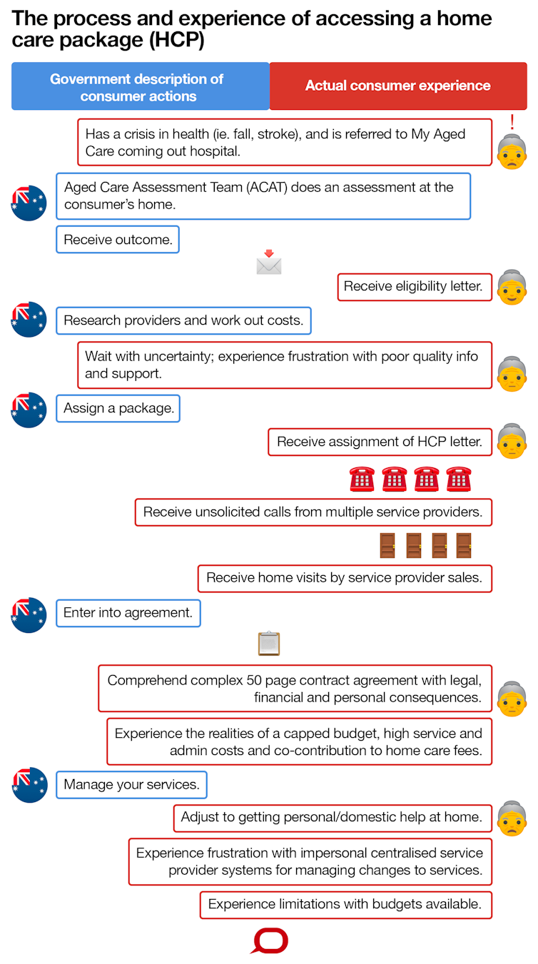
From July 1 2015, all home care packages have been delivered on what’s called a Consumer Directed Care basis. This means that, theoretically, home care providers must work with consumers to design and deliver services that meet their goals and care needs, as determined by an Aged Care Assessment Team. However, in reviewing the active steps outlined in the government pathway to access a package, we must consider the person who is navigating this path. They are frail older people with complex support needs, often seeking help at times of crisis. These include the growing number of older Australians living with multiple medical conditions and complex age-related syndromes such as dementia. After a person has been assessed, they will receive a letter informing them they are eligible. However, due to long waiting lists, this does not provide them with immediate access to care; most wait many months before they are actually assigned a package by My Aged Care. When they eventually receive a letter confirming their package, the consumer will be approached by various service providers. They will need to sign a complex contract with their chosen provider. If the consumer is feeling frustrated and confused during these early stages, this is only the beginning. The recent marketisation of home care means managing their own care requires going through impersonal, centralised provider systems.
The Conversation, CC BY-ND

People need clear information to choose a provider

The first thing people assigned a home care package need to do is choose a care provider. There are now close to 900 different providers offering home care packages. This includes not-for-profits, as well as a growing number of for-profit providers competing for new business.

In reality, however, few older people research different providers. Once they’re assigned a home care package, their name is placed on a centralised database accessible by all registered service providers. The person then receives unsolicited phone calls from the sales teams of different providers, offering their services and trying to make appointments to come and visit. For consumers, this represents a shift from a familiar government model of care provision to a market model. Research shows consumers often don’t understand consumer directed care, and this can leave them vulnerable to the forceful marketing tactics employed by some providers. It can also make negotiating a complex contract with legal, financial and personal implications very difficult. To make informed choices between providers, people need accessible information. There is currently insufficient information for older people and their families to compare services on indicators of quality (such as the number of complaints agencies receive, the training of staff, the types of specialist services they offer, and so on). To address this gap, the government must commit to collecting and publishing data on home care quality. This would drive service improvement and increase people’s ability to make informed choices between different providers.

Service and administrative fees

To make informed choices, people also need to be able to compare services on the basis of price. The average profit per client for home care package providers was A$2,832 in 2016-17, but there’s significant variability between providers’ fees. For example, the use of people’s individual care budgets to cover administration or case management fees ranges between 10-45% of their total package. High fees and administrative costs may reveal the profit-driven motives of a few unscrupulous providers. Because of administrative fees, many people are spending a high portion of their individual budgets on case management to support their care. While there’s evidence case management can provide clinical benefits for older people, in the context of the current home care funding model, it may also leave people with less money for direct care services than they need.

People need support to manage their packages

We’re currently looking at the experiences of people with dementia using home care packages. Unsurprisingly, we’re finding that while they are grateful for the services they’re receiving, they are having a difficult time managing their care. For some this may be due to their limited decision-making capacity, but for many, their choice and control is being limited as much by the service model. For example, to enable providers to compete in the open market, many have adopted central 1800 numbers to support people to manage their services. This means if consumers want to change something, they are funnelled through this system. Think about your own experience of service helplines, such as with telephone or energy companies. Now consider a woman with dementia who needs to call a 1800 number to change the time of her shower so she can see her doctor. Rather than communicating with a local and known case manager, she now needs to speak to someone she doesn’t know and who is not familiar with her care needs. Instead of facilitating choice and control, this demand on the consumer to constantly articulate their needs to unfamiliar people means many are frustrated, and some are even opting out of services.

How can we improve things?

The three words the government associates with consumer directed home care are choice, control and markets. But the system doesn’t foster control. Although consumers technically have choices, the marketised and bureaucratic approaches of service providers make it difficult for consumers to articulate and receive support for their personal choices. The processes, information and supports available to assist older people and their families are inadequate to facilitate the type of choices and control one might associate with “consumer directed” care. There’s an urgent need to improve the processes for accessing timely home care packages, particularly for those with complex support needs. This includes the quality and accessibility of information, resources and decision-making tools. There’s also a significant need for training, advocacy and impartial support for choice, particularly for people with limited decision-making capacity, such as those living with dementia. Research and practice in aged care and disability in other settings provide extensive resources for person-centred planning and decision making which could be adapted for use in our home care system.The Conversation Lyn Phillipson, NHMRC-ARC Dementia Development Fellow, University of Wollongong and Louisa Smith, Research Fellow at AHSRI, University of Wollongong This article is republished from The Conversation under a Creative Commons license. Read the original article.
Dr Linda Calabresi

To ablate, or not to ablate? That is the question. That’s what international researchers were investigating in two studies just published in The Journal of the American Medical Association. And the answer? As so often happens in medicine, the answer is: it depends. Looking at the two studies, patients with symptomatic atrial fibrillation had a greater improvement in their quality of life at the one year mark if they had undergone catheter ablation than if they had been treated with medical treatment alone. But not to diminish the importance of quality of life as a measure of success, other findings from the latest research are also worth noting. In the larger of the two studies, a randomised controlled controlled trial of over 2200  patients presenting with symptomatic AF, researchers found after four years of follow-up that there was no significant difference in mortality between the group who had received catheter ablation and those who were treated with drug therapy alone. Similarly, the rate of disabling stroke, serious bleeding and cardiac arrest were the same between the two groups. As one would expect there was a higher rate of AF recurrence among the drug therapy group as compared with the catheter ablation group (70% vs 50%), however that 50% recurrence rate among those who’d undergone the ablation procedure is still pretty high and overall among that intervention group 19.4% underwent a repeat procedure. But the study authors who came from 10 different countries did not seem too deflated by the result. While their study failed to show benefit for catheter ablation in any of the primary outcomes such as death or stroke they did find some advantage in terms of secondary outcomes, including quality of life. They also point to a trend toward benefit of the procedure even if that benefit wasn’t large enough to reach clinical significance. The other JAMA study involved just 155 patients who had symptomatic paroxysmal or persistent AF and who were randomised to receive either catheter ablation or drug therapy. The Scandinavian researchers were particularly assessing their symptoms and their quality of life. After four years, the catheter ablation group ‘produced 14% more patients who achieved complete or near complete relief from their AF symptoms.’ What’s more the quality of life improved for patients in both groups. However, the improvement was greater in the ablation group. So, what does it all mean? Firstly, it needs to be pointed out that, in keeping with the guidelines the majority of patients included in these trials were symptomatic – only 10% were asymptomatic. In other words, there have to be symptoms or another very good reason to consider ablation in a patient with AF. Secondly, overall, the ablation group was more successful than the drug therapy group in relieving those symptoms. As an accompanying editorial puts it: “For patients with symptoms, in whom quality of life is impaired by AF, catheter ablation can improve quality of life to a greater extent than drug therapy. However, patients who choose drug therapy will also likely experience significant improvements in quality of life and have no worse risk for the most concerning complications of AF, stroke and death. Thus, there is no mandate for these patients to undergo catheter ablation at this time.” And that’s where we’re at.

Reference:

Packer DL, Mark DB, Robb RA, Monahan KH, Bahnson TD, Poole JE, et al. Effect of Catheter Ablation vs Antiarrhythmic Drug Therapy on Mortality, Stroke, Bleeding, and Cardiac Arrest Among Patients With Atrial Fibrillation: The CABANA Randomized Clinical Trial. JAMA. 2019 Mar 15. DOI: 10.1001/jama.2019.0693 [Epub ahead of print] Mark DB, Anstrom KJ, Sheng S, Piccini JP, Baloch KN, Monahan KH, et al. Effect of Catheter Ablation vs Medical Therapy on Quality of Life Among Patients With Atrial Fibrillation: The CABANA Randomized Clinical Trial. JAMA. 2019 Mar 15. DOI: 10.1001/jama.2019.0692 [Epub ahead of print] Albert CM, Bhatt DL. Catheter Ablation for Atrial Fibrillation: Lessons Learned From CABANA. JAMA. 2019 Mar 15. DOI: 10.1001/jama.2018.17478 [Epub ahead of print] Blomström-Lundqvist C, Gizurarson S, Schwieler J, Jensen SM, Bergfeldt L, Kennebäck G, et al. Effect of Catheter Ablation vs Antiarrhythmic Medication on Quality of Life in Patients With Atrial Fibrillation: The CAPTAF Randomized Clinical Trial. JAMA. 2019 Mar 19; 321(11): 1059-68. DOI: 10.1001/jama.2019.0335
Jane Heller

With several hundred cases diagnosed each year, Australia has one of the highest rates of Q fever worldwide. Q fever is a bacterial infection which spreads from animals; mainly cattle, sheep and goats. It can present in different ways, but often causes severe flu-like symptoms. Importantly, the bacteria that cause Q fever favour dry, dusty conditions, and inhalation of contaminated dust is a common route of infection. There are now fears the ongoing droughts in Queensland and New South Wales may be increasing risk of the disease spreading. But there are measures those at risk can take to protect themselves, including vaccination.

What is Q fever and who is at risk?

Q fever is an infectious illness caused by the bacterium Coxiella burnetii, one of the most infectious organisms around. Q fever is zoonotic, meaning it can transmit to people from infected animals. It’s usually acquired through either direct animal contact or contact with contaminated areas where animals have been. Goats, sheep and cattle are the most commonly reported Q fever hosts, although a range of other animals may be carriers. Because of this association with livestock, farmers, abattoir workers, shearers, and veterinarians are thought to be at the highest risk of Q fever. People who also may be at risk include family members of livestock workers, people living or working near livestock transport routes, tannery workers, animal hunters, and even processors in cosmetics factories that use animal products. Q fever can be difficult to diagnose (it has sometimes been called “the quiet curse”). Infected people usually develop flu-like fevers, severe headaches and muscle or joint pain. These symptoms typically appear around two to three weeks after infection, and can last up to six weeks. A small proportion of people will develop persistent infections that begin showing up later (up to six years post-infection). These can include local infections in the heart or blood vessels, which may require lifelong treatment.

Are Q fever rates on the rise?

In Australia, 500 to 800 cases of Q fever (2.5 – 5 cases per 100,000 people) were reported each year in the 1990s according to the National Notifiable Diseases Surveillance System. A national Q fever management program was designed in 2001 to combat this burden. This program provided subsidised vaccination to at-risk people including abattoir workers, beef cattle farmers and families of those working on farms. Results were positive. Q fever cases decreased during the program and following its conclusion in 2006, leading to a historic low of 314 cases (1.5 cases per 100,000 people) in 2009. But since 2010, Q fever cases have gradually increased (558 cases or 2.3 per 100,000 were reported in 2016), suggesting further action may be necessary. Every year, the highest numbers of people diagnosed are from Queensland and NSW. And the true number of affected people is likely to be under-reported. Many infected people do not experience severe symptoms, and those who do may not seek health care or may be misdiagnosed.

Q fever and drought

The reason people are more susceptible to Q fever in droughts lies in the bacteria’s capacity to survive in the environment. Coxiella burnetii spores are very resilient and able to survive in soil or dust for many years. This also helps the bacteria spread: it can attach to dust and travel 10km or more on winds. The Q fever bacteria is resistant to dehydration and UV radiation, making Australia’s mostly dry climate a hospitable breeding ground. Hot and dry conditions may also lead to higher bacterial shedding rates for infected livestock. The ongoing drought could allow Q fever to spread and reach people who were previously not exposed. One study suggested drought conditions were probably the main reason for the increase in Q fever notifications in 2002 (there were 792 cases that year). This was the fourth driest year on record in Australia since 1900. We still need more evidence to conclusively link the two, but we think it’s likely that drought in Queensland and NSW has contributed to the increased prevalence of Q fever in recent years.

How can people protect themselves?

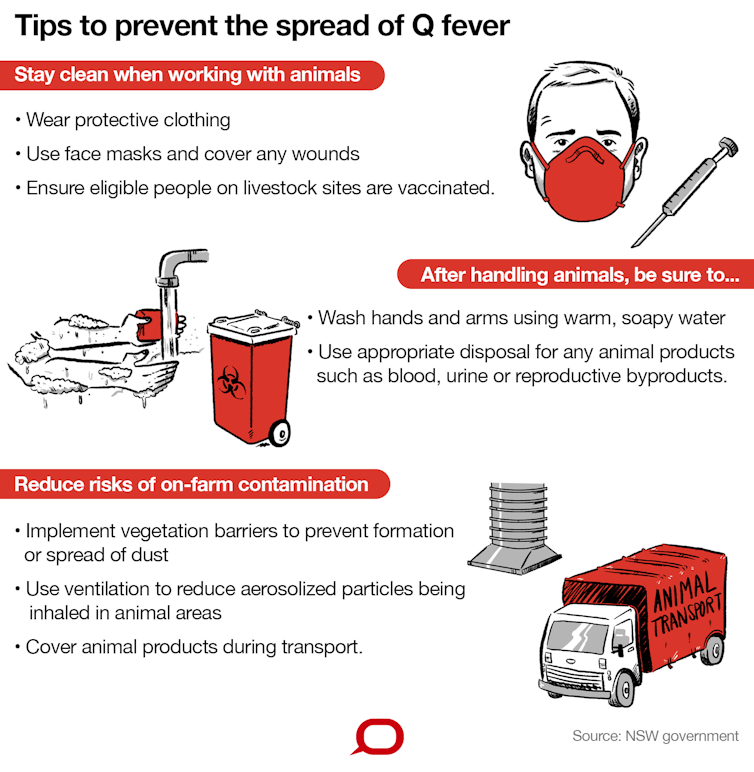
National guidelines for managing Q fever primarily recommend vaccination. The Q-VAX® vaccine has been in use since 1989. It’s safe and has an estimated success rate of 83–100%. However, people who have already been exposed to the bacteria are discouraged from having the vaccination, as they can develop a hypersensitive reaction to the vaccine. People aged under 15 years are also advised against the vaccine. Because the vaccine cannot be administered to everyone, people can take other steps to reduce risk. NSW Health recommends a series of precautions.
Author provided/The Conversation, CC BY-ND

What else can be done?

Vaccination for people in high-risk industries is effective to prevent Q fever infection, but must be administered well before people are actually at risk. Pre-testing requires both a skin test and blood test to ensure people who have already been exposed to the bacteria are not given the vaccine. This process takes one to two weeks before the vaccine can be administered, and it takes a further two weeks after vaccination to develop protection. This delay, along with the cost of vaccination, is sometimes seen as a barrier to its widespread use. Awareness of the vaccine may also be an issue. A recent study of Australians in metropolitan and regional centres found only 40% of people in groups for whom vaccination is recommended knew about the vaccine, and only 10% were vaccinated. We also need to better understand how transmission occurs in people who do not work with livestock (“non-traditional” exposure pathways) if we want to reduce Q fever rates.The Conversation Nicholas J Clark, Postdoctoral Fellow in Disease Ecology, The University of Queensland; Charles Caraguel, Senior lecturer, School of Animal and Veterinary Science, University of Adelaide; Jane Heller, Associate Professor in Veterinary Epidemiology and Public Health, Charles Sturt University; Ricardo J. Soares Magalhaes, Senior Lecturer Population Health & Biosecurity, The University of Queensland, and Simon Firestone, Academic, Veterinary Biosciences, University of Melbourne This article is republished from The Conversation under a Creative Commons license. Read the original article.
Dr Linda Calabresi

Got a patient with multiple sun spots on their head that need treatment? Well it looks like the old, tried and true 5FU cream is still the way to go, according to a randomised trial just published in the New England Journal of Medicine. Among more than 600 randomly assigned patients, Dutch researchers compared the effectiveness of four topical treatments commonly used to treat multiple actinic keratoses as part of a ‘field treatment’. In addition to the 5% fluorouracil cream (Efudix), the study looked at the effectiveness of 5% imiquimod cream (Aldara), methyl aminolevulinate photodynamic therapy (MAL PDT or Metvix PDT) and 0.015% ingenol mebutate gel (Picato gel). After 12 months, the study showed that the Efudix was the most effective in terms of maintaining a reduction of at least 75% of actinic keratoses from the baseline. In other words, this cream was the best of the four therapies, at getting rid of these sun spots completely. “And the differences between fluorouracil cream and imiquimod, PDT and ingenol mebutate were significant,” the study authors said. They found the likelihood of success for those patients using fluorouracil was almost 75%, compared with only 54% for imiquimod, 38% for PDT and 29% for those using ingenol mebutate. And this independent study didn’t do anything tricky with the dosing regimen either. “In our trial, we used the most commonly prescribed dosing regimens of the therapies studied,” they said. In terms of sticking to the dosing regimen, patients were much better adhering to the schedule when they were taking ingenol mubutate (99% adherence) or PDT (97%) rather than the fluorouracil (89%) or the imiquimod (88%), but this appeared to directly correlate with how often they had to take the therapy and for how long. Overall, however this adherence rate did not reflect treatment satisfaction rate. “Satisfaction with treatment and improvement in health-related quality of life at 12 months after the end of treatment were highest in the fluorouracil,” the study authors reported. Nothing like a treatment actually working to make a patient feel happy about having had it. A bonus of this study, according to the researchers was the inclusion of patients with the more severe actinic keratosis lesions (Grade III lesions), patients who have been commonly excluded from previous similar trials of topical treatments. “[Including these patients] is more representative of patients seen in daily practice,” they said. In addition to effectiveness, cost is another appealing factor for fluorouracil over the other treatments. This study has the capacity to change practice. The study authors quote the prevalence actinic keratoses among whites aged 50 and over as being at 37.5%. While cryotherapy remains the treatment of choice for single lesions, where there are multiple lesions present field treatment should be considered. Currently the guidelines for this field treatment don’t advocate one treatment over any other, more or less suggesting all four of the treatments in this study as being efficacious. However, as these Dutch researchers say “our results could affect treatment choices in both dermatology and primary care.”

Reference

Jansen MHE, Kessels JPHM, Nelemans PJ, Kouloubis N, Arits AHMM, van Pelt HPA, et al. Randomized Trial of Four Treatment Approaches for Actinic Keratosis. N Engl J Med. 2019 Mar 7; 380(10): 935-46.  DOI: 10.1056/NEJMoa1811850
Healthed

Cutaneous disorders are among the most common conditions presented to primary care doctors. Many are easily identifiable and may be dealt with effectively without the need for cutaneous biopsy. Nevertheless, in many instances the diagnosis is not obvious on clinical grounds. The rash may display atypical features or may not respond to therapy as predicted. In these cases, and when dealing with cutaneous tumours or worrying pigmented lesions, cutaneous biopsy with histological assessment becomes necessary. The art of cutaneous biopsy is to derive the maximum amount of information from the minimum amount of tissue, causing least discomfort to the patient. This will be achieved if due regard is given to the advantages and shortcomings of the various techniques available for biopsying cutaneous tissue, and if the pathologist is supplied with a good clinical history.

Clinical History

For several reasons, clinical history assists greatly in the interpretation of skin biopsies. Clinicopathological correlation is particularly important in many inflammatory cutaneous disorders. As the histological features can be very similar, clinical notes may help us to arrange a list of provisional diagnoses in order of likelihood. The key features to discuss with regard to cutaneous rashes include:
  • duration
  • distribution
  • description (macular, papular, vasculitic or vesicular)
  • drugs or other possible aetiological agents
  • provisional clinical diagnosis.
As there is wide variation in the normal microscopic picture from different sites, the area biopsied should also be stated. For biopsies performed to distinguish between squamous cell carcinoma and keratoacanthoma, the rate of growth of the lesion is important. When sending specimens of pigmented lesions, the degree of clinical suspicion should be stated, together with any history of melanoma within the individual or within the individual’s family. Any condition associated with cutaneous disorders, such as systemic lupus erythematosus, pregnancy or bone marrow transplant, should be mentioned in the clinical notes. The clinical history should also include the type of biopsy procedure used (see below) as this determines the way we handle the specimen in the laboratory. For example, the whole of an incisional biopsy will be blocked in order to gain the maximum amount of information, whereas an excisional biopsy will be transversely sectioned in order to fully assess the lateral excision margins in the case of a tumour biopsy.

Excision Biopsy

This is the best technique to use for pigmented lesions and cutaneous tumours. It allows for histological assessment and diagnosis of the lesion, and assessment of surgical excision margins. If appropriate, an orientation suture can be placed at one end of the excision, e.g. the superior end of the specimen, so that if the excision is inadequate, the margin involved can be indicated on an accompanying diagram. Occasionally, excision biopsy is appropriate for inflammatory cutaneous disorders where the condition is characterised by the formation of vesicles. The best chance of removing an intact vesicle (which greatly aids diagnosis) may be through excision.

Incision Biopsy

With incision biopsy, a thin elliptical biopsy is taken radially through the edge of the tumour or through the edge of a macular or annular rash. Incision biopsy is superior to punch biopsy for diagnosing rashes, more tissue is displayed on histological section and scarring is often reduced. A typical incision biopsy is 5-6 mm in length and about 2 mm in width. It should be deep enough to extend into the subcutaneous adipose tissue. The biopsy should run radially from the centre or central areas of the lesion to include approximately 1 mm of normal cutaneous tissue surrounding the lesion.

Punch Biopsy

Punch biopsies are easier to perform and, in general, are more convenient. Nevertheless, they nearly always yield less information than an incision biopsy. For tumours, the biopsy should be taken centrally. For cutaneous eruptions, the biopsy should be taken from an area typical of the rash. In some cases, multiple biopsies may increase the amount of information. In this procedure, it is best not to include normal skin. Punch biopsies come in various sizes. As 2 mm punches often yield inadequate information for diagnosis, a 3 mm punch biopsy is the smallest that should be used.

Shave Biopsy/Curettage

This technique is suitable for superficially-located lesions with plaque-like clinical features, e.g. seborrhoeic keratoses. It is not an appropriate technique for nodular lesions, cutaneous rashes or melanocytic lesions.

General comments concerning cutaneous biopsies

Preparation of the skin surface: Be gentle when cleaning the skin surface prior to biopsy; try not to disturb any overlying scale as the keratin layers sometimes contain diagnostic information (e.g. this is where dermatophytic fungi may be seen). Let any alcohol preparation dry before collecting specimens for immunofluorescence. Local anaesthesia: Only a small amount of local anaesthetic is required for punch biopsy procedures (0.5 mL maximum). Too much local anaesthetic within the tissues can distort the histological appearances and simulate dermal oedema. Marking the lesion: It is often prudent to mark the target area for biopsy with an ink marker, as some lesions can blanch following introduction of local anaesthetic. The erythema in many lesions is due to vascular dilatation occurring as part of the inflammatory disorder. Local anaesthetic can cause vasoconstriction and diminish the erythema clinically. This may result in a poorly targeted biopsy yielding subdiagnostic histology. Depth of biopsy: It is best to continue into the subcutaneous adipose tissue so that the entire dermis is represented on histological section. This helps greatly with the categorisation of many inflammatory skin disorders and also demonstrates the deep border of any cutaneous tumour. When performing a punch biopsy, the biopsy instrument appears to ‘give’ when it penetrates the dermal connective tissue into subcutaneous adipose tissue. A similar sensation will be noticed when dissecting free an incision biopsy. Care with biopsy tissue: All too often, after biopsy tissue has been retrieved from the patient, crush artefact occurs during its transfer into formalin. Crush artefact greatly distorts the histological appearance and repeat biopsy may become necessary. Rather than grasping the biopsy tissue with non-tooth forceps, it should be transferred to the specimen container using needle tips, a skin hook or fine forceps, delicately grasping one edge of the biopsy. Fixative: Ordinary blue, 10% buffered formalin supplied with the specimen jars is suitable for nearly all cutaneous biopsies, except those submitted for microbiological culture or immunofluorescent examination. Labelling: Please label all specimen containers with the patient’s name and details, which should match those stated on the request slip. Unlabelled specimens can still be processed and interpreted if they arrive with labelled paperwork; however, the medico-legal status of any generated report is doubtful. The report will usually be generated with a ‘specimen received unlabelled’ comment attached.

Conclusion

Diagnosing cutaneous conditions can be challenging. The chances of success are improved when the clinician, is armed with a variety of biopsy techniques for use in the correct clinical setting, and when the pathologist is supplied with an adequate clinical history.   - General Practice Pathology is a regular column each authored by an Australian expert pathologist on a topic of particular relevance and interest to practising GPs. The authors provide this editorial free of charge as part of an educational initiative developed and coordinated by Sonic Pathology.
Myriam Gharbi, Joseph H Drysdale, Hannah Lishman, Rosalind Goudie, Mariam Molokhia, Alan P Johnson, Alison H Holmes, Paul Aylin & Alastair D Hay

So here’s the exception that proves the rule. Urinary tract infections need immediate treatment with antibiotics to avoid an increased risk of sepsis and death. That’s the quite definitive conclusion from a large retrospective study involving GP data from the UK recently published in the BMJ. After analysing the records of over 150,000 patients, aged 65 and over presenting to their GP with a suspected or confirmed UTI, the researchers found those whose antibiotic treatment was delayed or deferred were up to eight times more likely to develop sepsis in the following 60 days compared to the group who were given antibiotics from the beginning. And those patients who were not given antibiotics at all, they were twice as likely to die as their medicated counterparts. Most of the infections were caused by Escherichia Coli, and trimethoprim or nitrofurantoin were the most common antibiotics prescribed. As the study showed, sepsis is not a common sequela of UTI, occurring in just .5% of cases. But the fact remains if antibiotics were delayed or withheld altogether the incidence jumped to 2.2% and 2.9% respectively which is significant and totally unnecessary. Understandably outcomes were worse the older the patient, and men had more adverse outcomes than women, but even accounting for multiple variable factors the basic conclusion remained the same. “Our study suggests the early initiation of antibiotics for UTI in older high-risk adult populations (especially men aged >85 years) should be recommended to prevent serious complications”, the study authors said. Of concern to the researchers was the relatively large number of older patients (about 7%) who were diagnosed with a UTI but not treated. They suggest antimicrobial stewardship programmes encouraging more judicious use of antibiotics may be at least, in part, to blame. That, and the risk of elderly patients developing Clostridium difficile infection following antibiotic use. But while ‘delayed or deferred’ antibiotic treatment was not generally associated with serious adverse outcomes for some self-limiting illnesses such as upper respiratory tract infections, this study suggests it is not a good idea for UTIs. “In our study, deferred antibiotics were associated with less severe adverse outcomes than no antibiotics for older adults but still showed a significantly higher risk of mortality compared with immediate antibiotics,” the researchers said. An accompanying editorial by a UK GP academic says the study highlights one of the many dilemmas that occur in general practice. “[GPs face] the daily challenge of ensuring that patients who are unlikely to benefit are not treated, whereas those who require antibiotics receive the right class, at the right time, at the right dose, and for the right duration,” he wrote. And while agreeing with the study authors concluding advice, that all older patients with suspected UTI should be treated from day one he does suggest further research is needed. Research could help determine the most appropriate antibiotic in this situation, and if there are any particular groups in this 65 and over cohort who it would be safe to leave off antibiotic treatment until the result of the culture and sensitivities are known. - Myriam Gharbi, NIHR Health Protection Research Unit, Imperial College London; Joseph H Drysdale, Department of Primary Care and Public Health, Imperial College London; Hannah Lishman, Medical School, St George's University of London UK; Rosalind Goudie, Nuffield Department of Population Health, University of Oxford, UK; Mariam Molokhia, Department of Primary Care and Public Health Sciences, King's College, London, UK; Alan P Johnson, Healthcare-Associated Infections and Antimicrobial Resistance Division, London, UK; Alison H Holmes, NIHR Health Protection Research Unit, Imperial College London; Paul Aylin, NIHR Health Protection Research Unit, Imperial College London This article is referenced from THEBMJ. Read the original article. - Alastair D Hay, Centre for Academic Primary Care, Bristol Medical School: Population Health Sciences, University of Bristol, Canynge Hall, Bristol, UK This article is referenced from THEBMJ. Read the original article.

Dr Cameron Webb

Western Australian health authorities recently issued warnings about Murray Valley encephalitis, a serious disease that can spread by the bite of an infected mosquito and cause inflammation of the brain. Thankfully, no human cases have been reported this wet season. The virus that causes the disease was detected in chickens in the Kimberley region. These “sentinel chickens” act as an early warning system for potential disease outbreaks.

What is Murray Valley encephalitis virus?

Murray Valley encephalitis virus is named after the Murray Valley in southeastern Australia. The virus was first isolated from patients who died from encephalitis during an outbreak there in 1951. The virus is a member of the Flavivirus family and is closely related to Japanese encephalitis virus, a major cause of encephalitis in Asia. Murray Valley encephalitis virus is found in northern Australia circulating between mosquitoes, especially Culex annulirostris, and water birds. Occasionally the virus spreads to southern regions, as mosquitoes come into contact with infected birds that have migrated from northern regions.

How serious is the illness?

After being transmitted by an infected mosquito, the virus incubates for around two weeks. Most people infected don’t develop symptoms. But, if you’re unlucky, you could develop symptoms ranging from fever and headache to paralysis, encephalitis and coma. Around 40% of people who develop symptoms won’t fully recover and about 25% die. Generally, one or two human cases are reported in Australia per year. Since the 1950s, there have been sporadic outbreaks of Murray Valley encephalitis, most notably in 1974 and 2011. The 1974 outbreak was Australia-wide, resulting in 58 cases and 12 deaths. It’s likely the virus has been causing disease since at least the early 1900s when epidemics of encephalitis were attributed to a mysterious illness called Australian X disease.

Early warning system

Given the severity of Murray Valley encephalitis, health authorities rely on early warning systems to guide their responses. One of the most valuable surveillance tools to date have been chooks because the virus circulates between birds and mosquitoes. Flocks of chickens are placed in areas with past evidence of virus circulation and where mosquitoes are buzzing about. Chickens are highly susceptible to infection so blood samples are routinely taken and analysed to determine evidence of virus infection. If a chicken tests positive, the virus has been active in an area. The good news is that even if the chickens have been bitten, they don’t get sick. Mosquitoes can also be collected in the field using a variety of traps. Captured mosquitoes are counted, grouped by species and tested to see if they’re carrying the virus. This method is very sensitive: it can identify as little as one infected mosquito in a group of 1,000. But processing is labour-intensive.

How can technology help track the virus?

Novel approaches are allowing scientists to more effectively detect viruses in mosquito populations. Mosquitoes feed on more than just blood. They also need a sugar fix from time to time, usually plant nectar. When they feed on sugary substances, they eject small amounts of virus in their saliva. This led researchers to develop traps that contain special cards coated in honey. When the mosquitoes feed on the cards, they spit out virus, which specific tests can then detect. We are also investigating whether mosquito poo could be used to enhance the sugar-based surveillance system. Mosquitoes spit only tiny amounts of virus, whereas they poo a lot (300 times more than they spit). This mosquito poo can contain a treasure trove of genetic material, including viruses. But we’re still working out the best way to collect the poo.

Staying safe from Murray Valley encephalitis

There is no vaccine or specific treatment for the virus. Avoiding mosquito bites is the only way to protect yourself from the virus. You can do this by:
  • wearing protective clothing when outdoors
  • avoiding being outdoors when the mosquitoes that transmit the virus are most active (dawn and dusk)
  • using repellents, mosquito coils, insect screens and mosquito nets
  • following public health advisories for your area.
The virus is very rare and your chances of contracting the disease are extremely low, but not being bitten is the best defence.The Conversation

- Ana Ramírez, PhD candidate, James Cook University; Andrew Francis van den Hurk, Medical Entomologist, The University of Queensland; Cameron Webb, Clinical Lecturer and Principal Hospital Scientist, University of Sydney, and Scott Ritchie, Professorial Research Fellow, James Cook University

This article is republished from The Conversation under a Creative Commons license. Read the original article.
Dr Linda Calabresi

Australian GPs are confident and competent at managing kids with bedwetting issues, new research confirms. But they are less sure what to do with children with daytime wetting or when childhood enuresis persists into adulthood, according to the study findings recently published in the Australian Journal of General Practice. As you may remember, back in late 2016, health professionals on the Healthed database were invited to participate in a survey designed by leading paediatric urologist, Dr Patrina Caldwell to investigate their knowledge and experience with managing urinary incontinence in childhood. Almost 1500 Australian health professionals responded, mostly GPs. Researchers found that 88% of survey participants reported being slightly or very knowledgeable about managing childhood urinary incontinence. Their confidence seems well-founded at least for nocturnal enuresis. Having been given multiple answer options about first-line management, 93% of participants correctly selected urotherapy and enuresis alarm training as the ideal first-line management for childhood enuresis. This is despite less than half (48%) being able to correctly identify the commonest cause of the condition as being abnormal physiology of sleep and bladder function. Over a third of people incorrectly thought childhood enuresis was simply a delay in developing toileting skills. However, it is a different kettle of fish when it comes to managing daytime urinary incontinence which only 81% of participants felt at least somewhat confident managing. Of concern was the finding that 18% of health professionals would treat this condition with inappropriate and potentially harmful treatments. More specifically, a small percentage of participants chose tricyclic antidepressants which used to be a popular treatment option but is now no longer recommended as first-line therapy for daytime urinary incontinence due to its potential side-effects. The situation was even worse for adult patients who had problems with enuresis that had persisted since childhood. Only 61% of participants felt they were even slightly knowledgeable managing these patients, although most chose the most appropriate first-line therapy of urotherapy and desmopressin. According to the study authors, the knowledge of the health professionals with regard to the various categories of urinary incontinence was largely reflective of the prevalence of each of the different conditions in their clinical experience. Most GPs were currently managing at least a few cases of nocturnal enuresis but the other two conditions were much rarer.

Reference

Caldwell PHY. Manocha R, Hamilton S, Scott KM, Barnes EH. Australian community health practitioners’ knowledge and experience with managing urinary incontinence that begins in childhood. Aust J Gen Pract. 2019 Jan; 48(1-2); 60-5. Available from: https://www1.racgp.org.au/ajgp/2019/january–february/managing-urinary-incontinence-that-begins-in-child
A/Prof Sanjaya Senanayake

The devastating Townsville floods have receded but the clean up is being complicated by the appearance of a serious bacterial infection known as melioidosis. One person has died from melioidosis and nine others have been diagnosed with the disease over the past week. The bacteria that causes the disease, Burkholderia pseudomallei, is a hardy bug that lives around 30cm deep in clay soil. Events that disturb the soil, such as heavy rains and floods, bring B. pseudomallei to the surface, where it can enter the body through through a small break in the skin (that a person may not even be aware of), or by other means. Melioidosis may cause an ulcer at that site, and from there, spread to multiple sites in the body via the bloodstream. Alternatively, the bacterium can be inhaled, after which it travels to the lungs, and again may spread via the bloodstream. Less commonly, it’s ingested. Melioidosis was first identified in the early 20th century among drug users in Myanmar. These days, cases tend to concentrate in Southeast Asia and the top end of northern Australia.

What are the symptoms?

Melioidosis can cause a variety of symptoms, but often presents as a non-specific flu-like illness with fever, headache, cough, shortness of breath, disorientation, and pain in the stomach, muscles or joints. People with underlying conditions that impair their immune system – such as diabetes, chronic kidney or lung disease, and alcohol use disorder – are more likely to become sick from the infection. The majority of healthy people infected by melioidosis won’t have any symptoms, but just because you’re healthy, doesn’t mean you’re immune: around 20% of people who become acutely ill with melioidosis have no identifiable risk factors. People typically become sick between one and 21 days after being infected. But in a minority of cases, this incubation period can be much longer, with one case occurring after 62 years.

How does it make you sick?

While most people who are sick with melioidosis will have an acute illness, lasting a short time, a small number can have a grumbling infection persisting for months. One of the most common manifestations of melioidosis is infection of the lungs (pneumonia), which can occur either via infection through the skin, or inhalation of B. pseudomallei. The challenges in treating this organism, though, arise from its ability to form large pockets of pus (abscesses) in virtually any part of the body. Abscesses can be harder to treat with antibiotics alone and may also require drainage by a surgeon or radiologist.

How is it treated?

Thankfully, a number of antibiotics can kill B. pseudomallei. Those recovering from the infection will need to take antibiotics for at least three months to cure it completely. If you think you might have melioidosis, seek medical attention immediately. A prompt clinical assessment will determine the level of care you need, and allow antibiotic therapy to be started in a timely manner. Your blood and any obviously infected body fluids (sputum, pus, and so on) will also be tested for B. pseudomallei or other pathogens that may be causing the illness. While cleaning up after these floods, make sure you wear gloves and boots to minimise the risk of infection through breaks in the skin. This especially applies to people at highest risk of developing melioidosis, namely those with diabetes, alcohol use disorder, chronic kidney disease, and lung disease.

Sanjaya Senanayake, Associate Professor of Medicine, Infectious Diseases Physician, Australian National University

This article is republished from The Conversation under a Creative Commons license. Read the original article.