Articles

Read the latest articles relevant to your clinical practice, including exclusive insights from Healthed surveys and polls.

By reading selected clinical articles, you earn CPD in the Educational Activities (EA) category whenever you click the “Claim CPD” button and follow the prompts. 

Dr Mike Armour

Many Australian women with endometriosis are reporting they’re being advised a reliable treatment or even possible cure for their endometriosis is to “go away and have a baby”. This message is consistent with what women from other countries are also being told by a wide range of sources from self-help books to web forums to medical professionals. Pregnancy as a natural cure for endometriosis appears to date back to the early 20th century. However, even into the 1950s and 1960s, when pregnancy was commonly recommended as a treatment for endometriosis, this evidence was based mostly on case reports of women whose endometriosis improved during pregnancy. Case reports are often unusual findings and don’t necessarily reflect what happens to most people. Pregnancy as a treatment for endometriosis does not appear in current international guidelines for the management of endometriosis. It’s also not mentioned as a treatment by Australian pelvic pain specialists and is classed as a “myth” by reputable endometriosis support sites.
Read more: Women with endometriosis need support, not judgement

Endometriosis and the lack of a cure

Endometriosis is the presence of tissue similar to the lining of the uterus outside the uterus itself. Accurate estimates of how many women in Australia have endometriosis are hard to find, but a common figure is around one in ten women during their reproductive years. While severe pain during the period is a common symptom of endometriosis, it’s so much more than just a “really bad period”. There’s almost no area of women’s lives that is not negatively affected by the condition. Current medical treatments, often using hormone therapy, are not always effective. And the side effects of many of the hormonal treatments can be particularly unpleasant for women, leading them to stop treatment. Excision surgery, in which the endometrial lesions are cut away, is the most effective current treatment. But surgery is not something most women enter into lightly, given the cost and risk of undergoing surgery. Unfortunately, even surgery is not always successful with around 50% of women having symptoms reoccur after five years. When we look at women around the world, it looks like having children does decrease the amount of period pain women have. A significant problem with this is we don’t know if these women had endometriosis, and these kinds of studies can’t tell us for sure if getting pregnant was responsible for this reduction in period pain.
Read more: Women aren’t responsible for endometriosis, nor should they be expected to cure themselves

Pregnancy, pain and the brain

Women with endometriosis, like other chronic pain conditions, have changes in the way their brains process pain. Nerves, especially in the pelvis, are also more sensitive than in women without chronic pain. The concept of “calming” these hyperactive pain pathways is an important treatment strategy in treating chronic endometriosis pain. Each time menstruation occurs it irritates these sensitive nerves and reinforces these pain pathways. One way to prevent this reinforcement of pain pathways can be by stopping regular menstruation entirely. This is a key reason women with endometriosis are so often treated with continuous use of hormonal contraceptives. During pregnancy there’s also a suppression of menstruation. So it’s possible during pregnancy there will be a reduction in endometriosis-related pain. It’s also just as possible pregnancy will make endometriosis-related pain worse, due to extra pressure on these sensitive pelvic nerves. We just don’t have the research to be able to answer this.
Read more - Health Check: are painful periods normal?
After giving birth, it’s quite possible the pain, if it had decreased, will return. This is especially true once women start having regular periods again, as there’s no evidence pregnancy shrinks endometrial lesions or changes pain processing in the long term, both major drivers of endometriosis pain.

Should pregnancy be recommended as a treatment?

Pregnancy might help reduce endometriosis symptoms, if only temporarily. But women with endometriosis often rightly feel upset and offended when advised to have a baby as a treatment strategy. There are also risks involved, as women with endometriosis are more likely to have pre-term births, increased rates of caesarean sections and an increased risk of miscarriage. Women shouldn’t have to bring another human into the world to relieve the pain of endometriosis. This is why we need to prioritise understanding the cause of endometriosis, finding effective treatments and eventually a cure.
The ConversationSyl Freedman, Co-founder EndoActive, M.A. Health Communication, MPhil (Medicine) Candidate, University of Sydney contributed to this article. Mike Armour, Post-doctoral research fellow in women's health,NICM, Western Sydney University This article was originally published on The Conversation. Read the original article.
Dr Linda Calabresi

Sometimes evidence proves what was long-suspected to be true. A new study, just published in JAMA Psychiatry shows women who took hormone replacement therapy early in the menopausal transition had almost half the risk of developing clinically significant depressive symptoms compared to women who took a placebo. The study also confirmed that women of this age and stage are at high risk of significant depression, with almost one third of women in the placebo group developing symptoms and signs of the condition over the 12 month study period. Previous research had suggested that hormone therapy could help manage existing depression in menopausal women, however according to the Canadian researchers, this study, conducted among initially euthymic women was the first to show hormone therapy’s role in preventing the affective disorder. More than 170 perimenopausal and early post-menopausal women were randomly assigned to receive transdermal oestradiol (0.1mg/day) and intermittent oral micronized progesterone or placebo patches and tablets for 12 months. They were assessed regularly for depression using a validated depression scale (CES-D). Women on placebo were more likely to record a score that equated with significant depression at least once over the study period (32.3%) compared with women taking the hormone therapy (17.3%). Interestingly, women who had had what the researchers called ‘stressful life events’ in the six months prior to enrolment in the trial actually had greater benefit from the hormone therapy. Whereas other possible confounders such as baseline vasomotor symptoms, a history of depression, and baseline oestradiol levels did not appear to affect the protective benefit of the therapy. The progesterone was given for 12 days every three months, to induce vaginal bleeding so the finding that this adverse effect was more common in the hormone therapy group was hardly surprising but of note the two groups did not differ in other adverse effects including headaches, bloating, breast tenderness, weight gain and GI symptoms. An accompanying editorial sounded a few warnings about the study including the fact that the oestrogen dose was higher than currently recommended for treating women with hot flushes and the progestin dose was less than that recommended to protect the endometrium. The two editorial authors, including Dr Martha Hickey, from the University of Melbourne also cautioned that using hormone therapy to prevent depression might result in prolonged hormone exposure with the known risks associated with this, and is not currently recommended for this indication. However, the study authors were cautiously enthusiastic about their findings saying, “If confirmed in a larger sample of early perimenopausal women, the findings of this study…suggest that hormone therapy may also be indicated for the prevention and/or treatment of depressive symptoms appearing in the early menopause transition, regardless of whether menopausal symptoms are present.” Ref: JAMA Psychiatry. doi:10.1001/jamapsychiatry.2017.3998

Dr Jenny Robson

Once specimens are received in the laboratory, microscopy is performed. An interim report is then released. Specimens are then set up on specialised agar containing antibiotics and cycloheximide to inhibit the growth of bacteria and saprophytic fungi. Cultures are incubated at 28°C for three weeks. If microscopy is positive (M+) and no pathogen (C-) has grown in the interim, specimens are held an extra week. Infrequently, where microscopy and culture of nail scrapings is negative and the diagnosis is still suspected, nails can be examined for fungal elements using special stains.

Results:

Specimen types have been subdivided into three anatomical categories (nails, hair and skin) based broadly on the three clinical presentations of onychomycosis, tinea capitis, and tinea corporis/ cruris/pedis. Onychomycosis refers to fungal infections of the nails and includes tinea unguium caused by dermatophytes but also non-dermatophyte fungi and yeasts, predominantly Candida spp.

Negative laboratory report:

A common reason for negative microscopy and /or culture is an incorrect clinical diagnosis. More than 50% of dystrophic nails do not have a fungal cause, so it is important to establish a correct laboratory diagnosis before treating a patient with an antifungal agent. Other reasons for false negative results include sampling variation associated with an inadequate specimen and/or splitting the sample to perform microscopy and culture; the presence of nonviable hyphae in the distal portion of the nail; uneven colonisation of the nail by fungus; and overgrowth by contaminant saprophytic fungi. Careful re-collection to obtain sufficient material may be necessary to confirm negative results.

Nails:

The analysis of nail specimens from the hands and feet. Fingernails: Of 1202 specimens processed, 59% were negative by both microscopy and culture; 11% had hyphae seen on microscopy but were negative by culture; 27% of all finger and thumbnail cultures grew a yeast, predominantly Candida albicans (88% of all positive nail/hand cultures). Only 3% of all fingernail specimens grew a dermatophyte.

Toenails:

57% of 5097 toenail cultures were negative by both microscopy and culture. 22% were positive by microscopy but culture negative for reasons stated previously. As the literature would suggest, yeast infection of toenails is rare. Dermatophytes (20%) predominate as the main cause of onychomycosis of the lower limbs. Transmission of these dermatophytes is usually via the feet and toe web spaces, which are the major reservoir on the human body. Onychomycosis can be regarded as the end stage of tinea pedis. Desquamated skin scales containing hyphae are shed and survive for months to years on floors and carpets. Infrequently non-dermatophyte moulds are implicated in toenail infections such as Aspergillus. There is some uncertainty as to the significance of these cultures, and repeat culture may be indicated.

Treatment options:

With tinea unguium, topical treatment is successful only with surgical removal of the nail combined with oral therapy. First-line treatment for all types of nail tinea consists of: 1. terbinafine (child < 20 kg: 62.5 mg; 20 to 40 kg: 125 mg) 250 mg orally, daily for six weeks for fingernails and 12 weeks for toenails or (if terbinafine is not tolerated) 2. itraconazole 200 mg orally, twice daily for seven days every month for 2-4 months or 3. fluconazole 150 to 450 mg orally, once weekly for 12 to 52 weeks. Successful management of candidiasis of the nail requires removal of risk factors e.g. water immersion.

Tinea capitis:

Of 414 hair samples submitted over this period, 329 (80%) were negative by both microscopy and culture. Dermatophytes isolated include M. canis (46%); T. tonsurans (42%); M. gypseum (5%); T. mentagrophytes (5%) and T. rubrum (2.5%). This condition afflicts predominantly prepubertal children. Clinically it can present as alopecia or a more inflammatory lesion (kerion). It is noteworthy that T. tonsurans, an anthropophilic fungus, is emerging as a common cause of tinea capitis in children and spreads easily from child to child.

Treatment options:

Tinea capitis often requires oral therapy to eradicate the infection. Treatment options include 1. griseofulvin fine particle (child: 20 mg/kg up to) 500 mg orally, daily for 4-8 weeks, or 2. terbinafine (child< 20 kg: 62.5 mg; 20 to 40 kg: 125 mg) 250 mg orally, daily for four weeks.

Tinea corporis/cruris/pedis:

Of the 7406 specimens received from skin sites, 73% were both microscopy and culture negative; 5% were positive only by microscopy. Of the 22% culture positive specimens, 19% grew a dermatophyte and 3% a yeast.

Treatment options:

When topical treatments have failed, recommended oral therapy includes: 1. griseofulvin fine particle (child: 10 to 20 mg/kg up to) 500 mg orally, daily for at least four weeks or 2. terbinafine (child <20 kg: 62.5 mg; 20 to 40 kg: 125mg) 250 mg orally, daily for at least two weeks, depending on the response or 3. itraconazole capsules 200 mg orally, twice daily for one week for tinea of the feet or hands or 4. itraconazole capsules 200 mg orally, once daily for one week for tinea elsewhere.
General Practice Pathology is a new regular column each authored by an Australian expert pathologist on a topic of particular relevance and interest to practising GPs. The authors provide this editorial, free of charge as part of an educational initiative developed and coordinated by Sonic Pathology.
Dr Jenny Robson

In Australia the commonest encounter with fungi in a medical sense is with superficial and cutaneous fungal infections such as those infecting the skin, scalp or nails.

Tinea or ringworm of the scalp, skin and nails

Fungal infection of the scalp (tinea capitus), skin (tinea) and nails (tinea unguium, onychomycosis) is usually caused by dermatophytes which have a unique ability to utilise keratin as a nutrient source due to the presence of the enzyme keratinase allowing colonisation of the stratum corneum. The presence of the fungus and its metabolic products can occasionally induce an allergic or inflammatory response in the host. The type and severity of the host response is often related to the species and strain of dermatophyte causing the infection. Following is a list of the dermatophytes that have been identified in our laboratories:
SPECIES NATURAL HABITAT INCIDENCE
Epidermophyton floccosum Humans Common
Trichophyton rubrum [worldwide] Humans Very common
Trichophyton interdigitale [anthropophilic] Humans Very common
Trichophyton tonsurans Humans Common
Trichophyton violaceum Humans Less common
Trichophyton concentricum Humans Rare
Trichophyton schoenleinii Humans Rare
Trichophyton soudanense Humans Rare
Trichophyton rubrum [African] Humans Rare
Microsporum audouinii Humans Less common
 
SPECIES NATURAL HABITAT INCIDENCE
Trichophyton interdigitale [zoophilic] Mice, rodents Common
Trichophyton erinacei Hedgehogs Rare
Microsporum canis Cats Common
 
SPECIES NATURAL HABITAT INCIDENCE
Microsporum gypseum Soil Common
Microsporum nanum Soil/pigs Rare
Microsporum cookei Soil Rare

Mycotic infections

Despite the majority of work done by mycology laboratories being concerned with superficial and cutaneous fungal infections, in recent years there has been an increase in fungal subcutaneous and systemic disease. Mycotic infections are usually classified according to the level of tissue involvement in the patient. The following table includes examples of such mycotic infections, their classification and an indication of their incidence.
LEVEL OF INFECTION MYCOSIS CAUSATIVE ORGANISM INCIDENCE
Superficial Pityriasis versicolor Seborrhoeic dermatitis including dandruff and follicular pityriasis Malassezia furfur (a lipophilic yeast) Common
Tinea nigra Hortaea werneckii Rare
White Piedra Trichosporon sp Common
Black Piedra Piedraia hortaea Rare
Cutaneous Dermatophytosis Trichophyton, Epidermophyton, Microsporum
Dermatomycosis Fungi other than dermatophytes
Candidiasis Candida species
Other yeasts Geotrichum, Trichosporon
Subcutaneous Sporotrichosis Sporothrix schenckii Rare
Chromoblastomycosis Fonsecaea, Phialophora, Cladophialophora etc Rare
Phaeohyphomycosis Cladophialophora, Exophiala, Bipolaris, Exserohilum, Curvularia Rare
Dimorphic Systemic Mycoses Histoplasmosis Histoplasma capsulatum Rare
Opportunistic Systemic Mycoses Candidiasis Candida albicans and related species Common
Cryptococcosis Cryptococcus neoformans Rare/Common
Aspergillosis Aspergillus fumigatus etc Rare

Specimen Collection

Laboratory diagnosis requires collection of an adequate amount of material for both microscopy and culture. The site needs to be cleaned with an alcohol wipe, which helps lower the contamination rate from bacteria, saprophytic moulds and yeasts that may overgrow a dermatophyte. As a health and safety precaution, scalpel blades should not be enclosed with specimen.

Scalp

In general, zoophilic fungi e.g. M.canis tend to cause ectothrix or involvement of the outside of the hair shaft and the lesions tend to be inflammatory, while anthropophilic fungi e.g. T.tonsurans result in endothrix or involvement of the hair shaft itself. The lesions are less inflammatory. Lustreless, broken, infected hairs should be sampled from the edge of a lesion. It is important to collect hair roots, as this is where fungal elements are detected. A scalpel blade may be used to dislodge crusts or scales in which hair stumps may be embedded.

Skin

Skin scrapings should be obtained by scraping the active border of the infection which usually is typically scaly, red and elevated and where the hyphae are present. In cases of kerion, swabs of the exudates should be collected. Separate specimens should be taken from the representative lesions on various parts of the body. When scraping feet, the site of most common involvement is between the fourth and fifth toes.

Nails

Scrape under the nail plate until the crumbling white degenerative portion is reached. All the keratin debris from under the nail should be collected directly onto a black collection card. The distal portion of the nail may need to be trimmed with nail clippers. Fungal elements remain viable for weeks at room temperature. Nail scrapings showing hyaline septate hyphae are diagnostic for a dermatophyte.

Transportation

Glass or plastic bottles with screw tops are not recommended since high relative humidity encourages proliferation of bacteria and reduces the chances of isolating fungi. Specimens for mycological studies are best submitted within the special black fungal scrapings cards provided by the laboratory or, if these are not available, within a folded paper (preferably dark) packet. Although delays are to be avoided, fungi are generally resistant to drying and survive transportation well at room temperature.

Negative Laboratory Report

The reasons for negative microscopy and/or culture include: • Incorrect clinical diagnosis • Sampling variation associated with an inadequate specimen • Splitting the sample to perform microscopy and culture • Presence of non-viable hyphae in the distal portion of nails • Uneven fungal colonisation of nails • Overgrowth by contaminant saprophytic fungi Careful recollection obtaining sufficient material may be necessary to confirm results. Adequate/Inadequate collection
General Practice Pathology is a new regular column each authored by an Australian expert pathologist on a topic of particular relevance and interest to practising GPs. The authors provide this editorial, free of charge as part of an educational initiative developed and coordinated by Sonic Pathology.
Dr Vivienne Miller

Based on an interview with Sydney dermatologist, Dr Rob Rosen conducted at the Annual Women’s and Children’s Health Update, Sydney in February 2018. Hyperhidrosis is a very distressing condition that equally affects both men and women, across all ethnicities.  It occurs in approximately 3% of the general population and the onset is in childhood or adolescence. However, only about one third of people affected seek medical advice. In addition to the physical effects, the psychological impact on affected individuals is significant. Embarrassment, anxiety and depression are very commonly associated with this condition. In primary hyperhidrosis, the sweat glands are normal but there is an apparently exaggerated sympathetic response. The sweat glands most susceptible to these sympathetic cholinergic effects are the eccrine glands found in the palms, soles and axillae. Primary, focal hyperhidrosis tends to be bilateral and symmetrical, occurring at least once a week. It usually commences before a person reaches their mid-twenties and is often familial.  In its typical form, and if there is nothing to suggest a secondary cause, it requires no investigations. Secondary hyperhidrosis is typically generalised, affecting the entire surface of the skin. By definition, it has an underlying cause, such as infection, endocrine disturbance, neuropathy, malignancy, menopause, drug withdrawal or the side-effect of medications.  A full history, examination and targeted investigations are required before this condition can be called idiopathic. “It is important to make the distinction between “generalised” and “focal” hyperhidrosis at the outset,” says Sydney dermatologist, Dr Rob Rosen The management of hyperhidrosis begins conservatively. By the time they present to a doctor they usually have already tried a range of antiperspirants.  Aluminium hydroxide 20%, topically, daily for four weeks should be trialled before further treatment is considered. Many patients develop localised irritation to this treatment, as it obstructs the eccrine ducts, causing their atrophy. The most effective management is Botulinum A toxin injections. This drug blocks the release of acetylcholine from presynaptic nerve terminals, thus inhibiting the stimulation of the eccrine sweat gland. The injections are done intradermally and retreatment is needed approximately every six months. Over time, this duration between treatments may become longer. Side effects include discomfort at the injection site and, less commonly, weakness of local muscles (especially relating to small muscles in the hand, for example, in palmer hyperhidrosis). In the research done by Dr Rosen t al, over 90% of patients were happy with this therapy.1 Oral anticholinergics such as oxybutynin (5mg to 15mg daily) or glycopyrrolate (1mg to 4mg daily) may be used and are most effective in refractory cases of generalised sweating. The anticholinergic side effects (urinary retention, dry mouth, constipation) tend to be a limiting factor in their use. Other treatments for hyperhidrosis tend to be either less effective or more invasive. For patients over 12 years old, there is a Medicare rebate for Botulinum A injection therapy for severe primary axillary hyperhidrosis, if aluminium hydroxide has failed and if it is administered by a dermatologist, neurologist or paediatrician. Some cosmetic clinics treat patients without a rebate and this is often a more expensive option.
  1. Rosen R, Stewart T. Results of a 10-year follow-up study of botulinum toxin A therapy for primary axillary hyperhidrosis in Australia. Intern Med J; 48;343-347.

Dr James Harraway

Non-Invasive Prenatal Testing (NIPT), the cell-free DNA-based blood test that screens for fetal chromosomal abnormalities, is fast becoming a routine part of obstetric care. NIPT at a glance During pregnancy, maternal plasma contains fragments of DNA from the mother and from the placenta (fetal DNA). The proportion of DNA fragments from particular chromosomes is usually very stable throughout pregnancy. If there is an excess of fetal fragments from one chromosome, the proportion of fragments from that chromosome will be changed. Inconclusive tests A key reason that NIPTs should precisely measures the amount of fetal DNA in the sample – the fetal fraction – is if there is insufficient fetal DNA, the result may merely reflect the genetic status of the mother. NIPT assays should report a result only if there is sufficient fetal DNA to be confident of accuracy. Rarely, a test for trisomy 21,18 and 13 cannot be reported. This occurs in 3% of women tested by Sonic Genetics and is usually because there is insufficient fetal DNA compared with maternal DNA in the mother’s plasma. This low fetal fraction can be due to a relative excess of maternal DNA and this can vary over time. It is more common in women with increased body weight, and more likely in the presence of infection and inflammation, or after exercise. It also occurs if the mother or fetus has some subtle benign variations in chromosome structure (copy number variants) that make estimating the proportion of fragments from a chromosome unreliable. In some instances, the DNA in the sample has degraded during collection and shipping to the laboratory, and the quality is insufficient for a reliable result. These factors interfere with quality control of the test. Two thirds of women will get a result on re-testing. However, if the second test is inconclusive, it should not be repeated. This occurs in 1% of pregnant women screened. It is also not worth using another form of non-invasive prenatal test. Other tests do not estimate the fetal fraction accurately and may provide false reassurance. A decision about other test modalities (combined first trimester screen, second trimester serum screen, detailed ultrasonography or invasive genetic testing such as CVS/amniocentesis) should be based on assessment of all identified risk factors and may require specialist consultation. More rarely (in 0.5 –1% of women) the test reports a result for trisomy 21, 18 and 13 but not for fetal gender and sex chromosome abnormalities. It is unlikely that a repeat test will provide a result. A decision about using fetal ultrasound or invasive genetic testing to document fetal gender should be based on assessment of need and any identified risk factors.   General Practice Pathology is a new regular column each authored by an Australian expert pathologist on a topic of particular relevance and interest to practising GPs. The authors provide this editorial, free of charge as part of an educational initiative developed and coordinated by Sonic Pathology.

Dr Linda Calabresi

The value of omega-3 fatty acids has come under fire lately. But now a new systematic review suggests they might have benefits beyond the previous therapeutic targets of depression, cardiac health, eye health and arthritis. Researchers have found that omega-3 polyunsaturated fatty acids (PUFA) might reduce the symptoms of clinical anxiety, particularly among those people who had a specific clinical condition be it medical (such as Parkinson disease) or psychological (premenstrual syndrome). “This systematic review…provides the first meta-analytic evidence, to our knowledge, that omega-3 PUFA treatment may be associated with anxiety reduction, which might not only be due to a potential placebo effect, but also from some associations of treatment with reduced anxiety symptoms,” the review authors said in JAMA. The finding is likely to be welcome news for patients with this condition. Be it the potential side-effects of medications or the cost and accessibility of psychological therapy, patients with anxiety, especially those with comorbid medical conditions are keen for alternative or at least supplementary safe, evidence-based treatments for their symptoms. Previous research, in both human and animal studies had found that a lack of omega-3 PUFAs could induce various behavioural and neuropsychiatric disorders. What had not been shown was whether taking this supplement was effective in reducing the specific anxiety symptoms. The review involved an extensive literature search through a wide range of databases including PubMed and Cochrane looking for trials that had assessed the anxiolytic effects of these fatty acids in humans. In the end they found 19 trials that matched their eligibility criteria, which allowed researchers to analyse the effect of supplementation in just over 1200 participants and compare it with about 1000 matched controls who didn’t take the fatty acids. Overall, they found ‘there was a significantly better association of treatment with reduced anxiety symptoms in patients receiving omega-3 PUFA treatment than in those not receiving it.’ Subgroup analysis also showed that those taking at least 2000mg or more of the omega-3 PUFA treatment were more likely to have reduced anxiety. And somewhat surprisingly, those patients receiving supplements containing less than 60% EPA did better than those taking formulations with a greater concentration of EPA. The studies in the review included very different cohorts, and because of this and the limited number of studies included, the authors understandably say the results need to be interpreted with caution. However, while bigger, better studies are still needed to prove the benefit of omega-3 PUFAs in patients with clinical anxiety, this research certainly does suggest that higher dose formulations of less than 60% concentration of EPA might have a role as at least adjunctive treatment to standard therapy.   Reference: Su KP, Tseng PT, Lin PY, Okubo R, Chen TY, Chen YW, et al. Association of Use of Omega-3 Polyunsaturated Fatty Acids With Changes in Severity of Anxiety Symptoms; A Systematic Review and Meta-analysis. JAMA Network Open [Internet]. 2018 Sep; 1(5): e182327. Available from: https://jamanetwork.com/journals/jamanetworkopen/fullarticle/2701735 doi:10.1001/jamanetworkopen.2018.2327.

Prof Andrew Whitehouse

New Australian autism guidelines, released today, aim to provide a nationally consistent and rigorous standard for how children and adults are assessed and diagnosed with autism, bringing to an end the different processes that currently exist across the country. There is no established biological marker for all people on the autism spectrum, so diagnosis is not a straightforward task. A diagnosis is based on a clinical judgement of whether a person has autism symptoms, such as social and communication difficulties, and repetitive behaviours and restricted interests. This is an inherently subjective task that depends on the skill and experience of the clinician. This judgement is made even more difficult by the wide variability in symptoms, and the considerable overlap with a range of other developmental conditions such as attention deficit/hyperactivity disorder (ADHD), intellectual disability, and developmental language disorder. Further complicating autism diagnosis in Australia is the lack of consistent diagnostic practices both within and between states and territories. This leads to patchy and inconsistent rules around who can access public support services, and the types of services that are available. It is not uncommon in Australia for a child to receive a diagnosis in the preschool years via the health system, for instance, but then require a further diagnostic assessment when they enter the education system. This is a bewildering situation that has a significant impact on the finite financial and emotional resources of families and the state. The new guidelines aim to address these inconsistencies and help people with autism and their families better navigate state-based support services. It also brings them into line with the principles of the National Disability Insurance Scheme (NDIS), which seeks to determine support based on need rather than just a diagnosis.

National guidelines

In June 2016, the National Disability Insurance Agency (NDIA) and the Cooperative Research Centre for Living with Autism (Autism CRC), where I’m chief research officer, responded to these challenges by commissioning the development of Australia’s first national guidelines for autism assessment and diagnosis. We undertook a two-year project that included wide-ranging consultation and extensive research to assess the evidence. The guidelines do not define what behaviours an individual must show to be diagnosed with autism. These are already presented in international manuals, such as the American Psychiatric Association’s Diagnostic and Statistical Manual (DSM-5) and the World Health Organisation’s International Classification of Diseases (ICD-11). What the new guidelines provide is a detailed description of the information that needs to be collected during a clinical assessment and how this information can be used to inform the ongoing support of that person, including through a diagnosis of autism. The guidelines include 70 recommendations describing the optimal process for the assessment and diagnosis of autism in Australia.

Understanding strengths and challenges

A diagnostic assessment is not simply about determining whether a person does or doesn’t meet criteria for autism. Of equal importance is gaining an understanding about the key strengths, challenges and needs of the person. This will inform their future clinical care and how services are delivered. In essence, optimal clinical care is not just about asking “what” diagnosis an individual may have, but also understanding “who” they are and what’s important to their quality of life. We know diagnosis of autism alone is not a sound basis on which to make decisions about eligibility for support services such as the NDIS and state-based health, education and social support systems. Some people who meet the diagnostic criteria for autism will have minimal support needs, while other individuals will have significant and urgent needs for support and treatment services but will not meet diagnostic criteria for autism at the time of assessment. Some people may have an intellectual disability, for example, but not show the full range of behaviours that we use to diagnose autism. Others may present with the latter, but not the former. In the context of neurodevelopmental conditions such as autism, it is crucial that a persons’s needs – not the presence or absence of a diagnostic label – are used to determine eligibility and prioritisation of access to support services.

What may influence an autism assessment?

The guidelines also detail individual characteristics that may influence the presentation of autism symptoms. Gender is one key characteristic. Males are more commonly diagnosed with autism than females. But there is increasing evidence that autism behaviours may be different in males and females. Females may be better able to “camouflage” their symptoms by using compensatory strategies to “manage” communication and social difficulties. It is similarly important to consider the age of the person being assessed, because the presentation of autism symptoms changes during life. The guidelines provide information on how gender and age affect the behavioural symptoms of autism. This will ensure clinicians understand the full breadth of autistic behaviours and can perform an accurate assessment. The next step is for all clinicians and autism service providers across Australia to adopt and implement the guidelines. This will ensure every child and adult with autism can receive the optimal care and support.
Daryl Efron and Harriet Hiscock

The rate of medications dispensed for attention-deficit hyperactivity disorder (ADHD) in children aged 17 and under increased by 30% between 2013-14 and 2016-17. The Australian Atlas for Healthcare Variation, released today, shows around 14,000 prescriptions were dispensed per 100,000 children aged 17 and under in 2016-17, compared with around 11,000 in 2013-14. The atlas for 2016-17 also showed some areas had a high dispensing rate of around 34,000 per 100,000 while the area with the lowest rate was around 2,000 per 100,000 – a 17-fold difference. This difference is much lower than in 2013-14, when the highest rate was 75 times the lowest. For decades people have been concerned too many children could be diagnosed with ADHD and treated with medications. We are conducting a study called the Children’s Attention Project, following 500 children recruited through Melbourne schools. So far, we have found only one in four children who met full ADHD criteria were taking medication at age ten. So it looks like, if anything, more children with ADHD should be referred for assessment and consideration of management options.

How many kids are medicated?

ADHD is the most common neurodevelopmental disorder of childhood – the prevalence is around 5% in Australia. Children with ADHD have great difficulty staying focused, are easily distracted and have poor self-control. Many are also physically hyperactive, especially when they are young. To be diagnosed, children need to have major problems from their ADHD symptoms both at home and school. These include learning difficulties, behavioural problems and trouble making friends. Young people with ADHD are more likely to fail school, have lower quality of life, experience substance abuse issues and teenage pregnancy, or end up in prison. Medication can make a big difference to these children’s lives. While there are many ways to help children with ADHD, stimulant medication is the most effective treatment. All international clinical guidelines recommend it for children with significant ADHD that persists after non-medication approaches have been offered. Our previous research found that about 80% of children diagnosed with ADHD by a paediatrician (the main medical specialty that manages ADHD) in Australia are treated with medication. The atlas shows the proportion of children and adolescents who had at least one ADHD medication prescription dispensed was 1.5% in 2013-14 and 1.9% in 2016-7. This is similar to the prevalence of stimulant medication prescription in previous Australian studies in the past 15 years. It sits between the US (high) and Europe (low) and is not excessive given the prevalence of the condition. The Children’s Attention Project found those with the most severe symptoms were more likely to be prescribed medications, as were those from families of lower socioeconomic status. Other Australian studies have found similar results. This is not surprising as ADHD does appear to be more common in children from socioeconomically disadvantaged families. Our research suggests that disadvantaged families in Australia appear to be able to access services for ADHD, at least in metropolitan centres.

Why does it vary between areas?

The atlas finding that there is considerable regional variation in prescribing of stimulant medications in Australia has been identified in previous studies and needs to be better understood. Some variation in health care is normal and good, but too much suggests there may be a problem with the quality of care or access to care. For example, greater prescribing in regional areas may reflect lack of timely access to non-pharmacological services. We do need to keep watching this space, monitoring rates and regional variation of medication use. A landmark study in the US, published in 1999, compared medication with intensive parent and teacher behaviour training. The children who received medication had a much greater reduction in ADHD symptoms. But medication is only one consideration in ADHD. Other supports are also important. Behavioural therapies can help reduce anxiety and behaviour problems in children with ADHD and improve relationships with parents and teachers. However, accessing psychologists can be hard for many families. While Medicare rebates are available for up to ten sessions per year, costs can still be a barrier. In our research, Victorian parents reported out-of-pocket costs of up to A$200 per session with a psychologist. ADHD is not considered a disability under the National Disability Insurance Scheme, so families are not eligible for funding packages. Further research is needed to better understand the factors influencing access to care for Australian children with ADHD, and why there is such variation in rates of prescribing between regions. We also need to ensure children across Australia get equitable access to non-medication management. We need evidence-based clinical guidelines relevant to the Australian healthcare system, which is quite different from places such as the UK and US. This work must include adult ADHD, which is an emerging area with a raft of clinical and service system complexities.The Conversation This article is republished from The Conversation under a Creative Commons license. Read the original article.
Lesley McCowan

A New Zealand-led international study published today provides the strongest evidence yet that women can more than halve their risk of stillbirth by going to sleep on either side during the last three months of pregnancy. This mega study (known as individual participant data meta-analysis) has also confirmed the risk of stillbirth associated with sleeping on the back applies to all pregnant women in the last trimester of pregnancy.

Risk factors

In New Zealand, stillbirth is defined as the loss of a baby after 20 weeks of pregnancy. An estimated 2.64 million babies die before birth globally each year, and around 300 babies are stillborn in Aotearoa New Zealand each year. About one in every 500 women in New Zealand will experience the tragedy of a late stillbirth and lose their baby during or after 28 weeks of pregnancy. We have analysed all available data worldwide from five previous studies, including our earlier research, the 2011 Auckland Stillbirth Study, which first identified a link between mothers’ sleeping position and stillbirth risk. The main finding in the mega study, which included information from 851 bereaved mothers and 2,257 women with ongoing pregnancies, was that going to sleep lying on the back (supine) from 28 weeks of pregnancy increased the risk of stillbirth 2.6 times. This heightened risk occurred regardless of the other known risk factors for stillbirth. However, the risk is additive, meaning that going to sleep on the back adds to other stillbirth risk factors, for example, a baby who is growing poorly in the womb. Existing common risk factors for late stillbirth are not easily modifiable. They include advanced maternal age (over 40), obesity, continued cigarette smoking and an unborn baby that is growing poorly, especially if the poor growth is not recognised before birth. Women also have a higher risk during their first pregnancy, or if they have already had three or more babies. Women of Pacific and South Asian ethnicity also have an elevated risk of late stillbirth, compared with European women. If modifiable risk factors can be identified, some of these baby deaths could be prevented. Importantly, our mega study has shown that if every pregnant woman went to sleep lying on her side after 28 weeks of pregnancy, approximately 6% of late stillbirths could be prevented. This could save the lives of about 153,000 babies each year worldwide.

Reduced blood flow

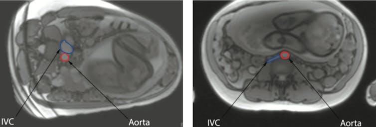
The relationship between the mother going to sleep lying on her back and stillbirth is biologically plausible. A supine position in late pregnancy is associated with reduced blood flow to the womb. Hence, women in labour and women having a caesarean section are routinely tilted onto their side to improve blood supply to the baby. Recent research carried out at the University of Auckland has provided sophisticated evidence about how the mothers’ position influences blood flow. Results obtained using Magnetic Resonance Imaging (MRI) demonstrate the major vessel in the mother’s abdomen, the inferior vena cava, being compressed by the pregnant womb when she is lying on her back. This reduces flow through this vessel by 80%.
The MRI images show the inferior vena cava (IVC) in blue and the aorta in red. In the left image, the mother is lying on her left side, while in the right image, she is on her back. provided, CC BY-SA
Although the mother’s circulation responds by increasing the flow through other veins, this does not fully compensate. The mother’s aorta, the main artery which carries oxygen-rich blood from her heart, is also partly compressed when the mother lies on her back. This decreases blood flow to the pregnant uterus, placenta and baby. We speculate that while healthy unborn babies can compensate for the reduced blood supply, babies that are unwell or vulnerable for some other reason may not cope. For example, our mega study showed that the risk of stillbirth after 28 weeks of pregnancy is increased approximately 16 times if a mother goes to sleep lying on her back and also is pregnant with a very small baby.

What to do

New Zealand research has shown that pregnant women can change their sleeping position. In a recent survey conducted in pregnant women from south Auckland, a community that has a high rate of stillbirth, more than 80% of women surveyed stated that they could change the position they went to sleep in with little difficulty if it was best for their baby. Our advice to pregnant women from 28 weeks of pregnancy is to settle to sleep on their side to reduce the risk of stillbirth, and to start every sleep, including day-time naps, on the side. It does not matter which side. It is common to wake up on the back, but we recommend that if this happens, women should simply roll back on to either side.The Conversation Lesley McCowan, Professor, Obstetrics & Gynecology, University of Auckland and Robin Cronin, Midwife researcher, University of Auckland This article is republished from The Conversation under a Creative Commons license. Read the original article.
Dr Edmond Chan

  “We don’t have to live in fear anymore.” That’s the common refrain from hundreds of parents of preschoolers with peanut allergy that my colleagues and I have successfully treated with peanut “oral immunotherapy” over the past two years. Oral immunotherapy (OIT) is a treatment in which a patient consumes small amounts of an allergenic food, such as peanut, with the dose gradually increased to a target maximum (or maintenance) amount. The goal for most parents is to achieve desensitization — so their child can ingest more of the food without triggering a dangerous reaction, protecting them against accidental exposure. A recent study published in The Lancet has suggested that this treatment may make things worse for children with peanut allergies. The researchers behind the meta-analysis argue that children with peanut allergies should avoid peanuts. This study has limitations however. It did not include a single child under the age of five years old. And it runs the risk of confusing parents. My colleagues and I have seen firsthand that oral immunotherapy is not only safe, but is well tolerated in a large group of preschool children. We published data demonstrating this recently in the Journal of Allergy and Clinical Immunology: In Practice.  

Safe for preschoolers

For any parent of a child with severe allergy, the idea of giving them even a small amount of the allergenic food might give them pause. I don’t blame them — giving a child a known allergen is a daunting thought. Some allergists share this fear and do not offer OIT to patients in their clinics due to safety concerns. To assess the safety of oral immunotherapy, we followed 270 children across Canada between the ages of nine months and five years who were diagnosed with peanut allergy by an allergist. The children were fed a peanut dose, in a hospital or clinic, that gradually increased at every visit. Parents also gave children the same daily dose at home, between clinic visits, until they reached the maintenance dose. We found that 243 children (90 per cent) reached the maintenance stage successfully. Only 0.4 per cent of children experienced a severe allergic reaction. Out of over 40,000 peanut doses, only 12 went on to receive epinephrine (0.03 per cent). Our research provides the first real-world data that OIT is safe for preschool-aged children with peanut allergy when offered as routine treatment in a hospital or clinic, rather than within a clinical trial.  

The Lancet study was of older children

So why does the meta-analysis published in The Lancet show that peanut OIT increases allergic reactions, compared with avoidance or placebo? The researchers behind this study argue that avoidance of peanut is best for children with peanut allergy. They describe that in older children, the risk of anaphylaxis is 22.2 per cent and the risk of serious adverse events is 11.9 per cent. It is important for parents to note that The Lancet study only assessed children aged five and older participating in clinical trials (average age nine years old), and the researchers don’t even mention this as a limitation of their analysis. Our study, on the other hand, assessed preschool children (average age just under two years old) in the real world outside of research. While I agree that there are certainly more safety concerns in older children, and more research is needed to see which of them would most benefit, our results demonstrate with real-world data that, in preschoolers, OIT is a game-changer.  

For many patients, benefits outweigh risks

It isn’t rocket science that avoiding what one is allergic to will be safer than eating it. An analogy is knee replacement surgery. Of course, not having knee replacement surgery would be “safer” than having the surgery. But not having knee replacement surgery doesn’t provide any potential of benefits and also provides little hope for families. Likewise, telling parents of children with peanut allergy that avoidance is the only option outside research fails to take into account the negative long-term consequences of avoidance — such as poor quality of life, social isolation and anxiety. Allergists and the medical community as a whole must stop confusing parents with endless mixed messages about OIT both within and outside of research. The fact is, many allergists are already offering OIT outside of research. In our current era of basing medical treatment decisions on a comparison of risks versus benefits, there is simply no one-size-fits-all approach. Rather than concluding that all children with peanut allergy should be managed with avoidance, we should be concluding that there are some patients, such as preschoolers, for whom the benefits of offering this treatment outweigh the risks. OIT has proven to be effective in many studies, and we will similarly follow the progress of our patients long term to track effectiveness. The bottom line is this: OIT is safe for preschool children and should be considered for families of those very young children with peanut allergy who ask for it.The Conversation  

- Edmond Chan, Pediatric Allergist; Head & Clinical Associate Professor, Division of Allergy & Immunology, Department of Pediatrics, Faculty of Medicine; Investigator, BC Children's Hospital Research Institute, University of British Columbia

This article is republished from The Conversation under a Creative Commons license. Read the original article.
Dr Mihiri Silva

If you imagine a teething child, what do you see? An irritable tot with a fever, in pain, and generally unwell? Teething’s a normal developmental process that people have long associated with illness. However, the evidence says otherwise. How strong is this evidence? Is there anything you can do to help a teething child? What about teething gels and teething necklaces? Teething is when new teeth emerge through the gums, and usually starts at about six months of age. A review of 16 studies found that although teething was linked with signs and symptoms, these were usually mild involving gum irritation, irritability, and drooling. Although body temperature may be slightly raised, the review found poor evidence to suggest teething caused fever. Many symptoms linked to teething, like irritability, sleep disturbance and drooling, are difficult to measure objectively and are based on what parents report, which is subjective and may be inaccurate. And, as teething comes and goes, and its timing is relatively unpredictable, recording even measurable symptoms like temperature changes in a reproducible, reliable way is virtually impossible. So teething problems seem to be over-reported in the types of studies that rely on people remembering what happened.

What else could cause the symptoms?

Other biological triggers may in fact explain the symptoms traditionally linked to teething. Teething coincides with normal changes in children’s immunity; the mother’s antibodies are transferred to babies in pregnancy and help protect the baby in the first 6-12 months of life, but start to wane at about the same time as teething. This, together with behavioural changes as infants start to explore their surroundings, increases the chances of catching viral infections with symptoms like those reported for teething. Separation anxiety and normal changes in sleep patterns may also account for irritability and sleep disturbances, which may be mistakenly attributed to teething. As teething symptoms are generally likely to be mild and focused on the mouth, parents are warned against presuming that signs of illness in other parts of the body are due to teething. That’s because this may delay the detection of potentially serious infections that may need medical attention. It may also delay parents getting help settling their child to sleep.

How about teething gels?

The search for solutions to the perceived problem of teething may lead parents to pin their hopes on gels, toys and other products, none of which have been scientifically assessed to alleviate teething symptoms. Nevertheless, teething gels usually contain a variety of ingredients that help relieve supposed teething-related symptoms. Some, such as the recently discontinued Adelaide Women’s and Children’s Hospital Teething Gel, contain the anaesthetic lidocaine. Very little lidocaine is absorbed into the body when applied to the gums, and only minor complications like vomiting have been reported in Australia. However, accidental swallowing and applying too much can lead to poisoning, resulting in seizures, brain injury, and heart problems. The decision to discontinue the gel follows a 2014 warning issued by the US Food and Drug Administration against using teething gels with topical anaesthetics, after reports of infant and child hospitalisation and death. There have also been warnings about teething gels containing benzocaine. This is another anaesthetic applied to the gums that can lead to a dangerous and fatal blood condition called methaemoglobinaemia, which affects the blood’s ability to carry oxygen. Another common ingredient in popular teething gels is choline salicylate, an anti-inflammatory similar to aspirin. This increases the risk of liver disease and brain injury if the child eats too much. This may also carry the risk of Reye syndrome, a rare but serious condition that can lead to seizures, loss of consciousness and death. Reye syndrome has been linked to the use of aspirin in children, particularly during viral infections. A case of suspected teething gel-induced Reye syndrome in 2008 led to the products being contraindicated (warned against) in children in the UK. A number of young Australian children who used too much salicylate-containing teething gel have also reportedly been hospitalised with side-effects. But the products are still available in Australia.

How about ‘natural’ products?

Although a range of “natural” and homeopathic teething solutions are heavily marketed to parents of young children, these too have risks. A manufacturer recently recalled a range of natural teething gels after cases of reported poisoning. And US regulatory authorities found the same range contained higher than reported levels of belladonna, a poisonous plant that despite its dangers is used as a homeopathic pain killer and sedative. In searching for “natural” therapies, parents are also turning to amber teething necklaces that supposedly relieve teething symptoms. Amber is a fossilised tree resin that has historically been suggested to have anti-inflammatory properties. However, several widely reported cases of strangulation have led to warnings from both US and Australian regulatory authorities. There is currently no scientific evidence these necklaces work. The Australian Competition and Consumer Commission (ACCC) says amber and other “teething” necklaces, even when mothers wear them, are:
…colourful and playful in design, and may be confused with toys.
All toys for children aged 36 months and below, including teething toys, are strictly regulated by Australian standards. As the ACCC warns, teething necklaces are unlikely to fulfil this requirement.

What to do?

So what are the best options to relieve teething symptoms? With a lack of any good-quality evidence to recommend any specific therapy, experts suggest the best remedy is affection and attention. Rubbing a clean finger on the gum, or applying gentle, firm pressure with a cooled (but not frozen), clean washcloth or teething ring may provide some relief. Although it’s hard to know exactly how these work, they are unlikely to lead to serious problems. Teething can be a difficult time, but it will eventually pass. In the meantime, it is important that parents avoid falling prey to supposed cures that are not only unproven, but are also potentially dangerous. Mihiri Silva, Paediatric dentist, Senior Lecturer and Post-doctoral Research Fellow, Murdoch Children's Research Institute This article is republished from The Conversation under a Creative Commons license. Read the original article.