Articles

Read the latest articles relevant to your clinical practice, including exclusive insights from Healthed surveys and polls.

By reading selected clinical articles, you earn CPD in the Educational Activities (EA) category whenever you click the “Claim CPD” button and follow the prompts. 
Jane Heller

With several hundred cases diagnosed each year, Australia has one of the highest rates of Q fever worldwide. Q fever is a bacterial infection which spreads from animals; mainly cattle, sheep and goats. It can present in different ways, but often causes severe flu-like symptoms. Importantly, the bacteria that cause Q fever favour dry, dusty conditions, and inhalation of contaminated dust is a common route of infection. There are now fears the ongoing droughts in Queensland and New South Wales may be increasing risk of the disease spreading. But there are measures those at risk can take to protect themselves, including vaccination.

What is Q fever and who is at risk?

Q fever is an infectious illness caused by the bacterium Coxiella burnetii, one of the most infectious organisms around. Q fever is zoonotic, meaning it can transmit to people from infected animals. It’s usually acquired through either direct animal contact or contact with contaminated areas where animals have been. Goats, sheep and cattle are the most commonly reported Q fever hosts, although a range of other animals may be carriers. Because of this association with livestock, farmers, abattoir workers, shearers, and veterinarians are thought to be at the highest risk of Q fever. People who also may be at risk include family members of livestock workers, people living or working near livestock transport routes, tannery workers, animal hunters, and even processors in cosmetics factories that use animal products. Q fever can be difficult to diagnose (it has sometimes been called “the quiet curse”). Infected people usually develop flu-like fevers, severe headaches and muscle or joint pain. These symptoms typically appear around two to three weeks after infection, and can last up to six weeks. A small proportion of people will develop persistent infections that begin showing up later (up to six years post-infection). These can include local infections in the heart or blood vessels, which may require lifelong treatment.

Are Q fever rates on the rise?

In Australia, 500 to 800 cases of Q fever (2.5 – 5 cases per 100,000 people) were reported each year in the 1990s according to the National Notifiable Diseases Surveillance System. A national Q fever management program was designed in 2001 to combat this burden. This program provided subsidised vaccination to at-risk people including abattoir workers, beef cattle farmers and families of those working on farms. Results were positive. Q fever cases decreased during the program and following its conclusion in 2006, leading to a historic low of 314 cases (1.5 cases per 100,000 people) in 2009. But since 2010, Q fever cases have gradually increased (558 cases or 2.3 per 100,000 were reported in 2016), suggesting further action may be necessary. Every year, the highest numbers of people diagnosed are from Queensland and NSW. And the true number of affected people is likely to be under-reported. Many infected people do not experience severe symptoms, and those who do may not seek health care or may be misdiagnosed.

Q fever and drought

The reason people are more susceptible to Q fever in droughts lies in the bacteria’s capacity to survive in the environment. Coxiella burnetii spores are very resilient and able to survive in soil or dust for many years. This also helps the bacteria spread: it can attach to dust and travel 10km or more on winds. The Q fever bacteria is resistant to dehydration and UV radiation, making Australia’s mostly dry climate a hospitable breeding ground. Hot and dry conditions may also lead to higher bacterial shedding rates for infected livestock. The ongoing drought could allow Q fever to spread and reach people who were previously not exposed. One study suggested drought conditions were probably the main reason for the increase in Q fever notifications in 2002 (there were 792 cases that year). This was the fourth driest year on record in Australia since 1900. We still need more evidence to conclusively link the two, but we think it’s likely that drought in Queensland and NSW has contributed to the increased prevalence of Q fever in recent years.

How can people protect themselves?

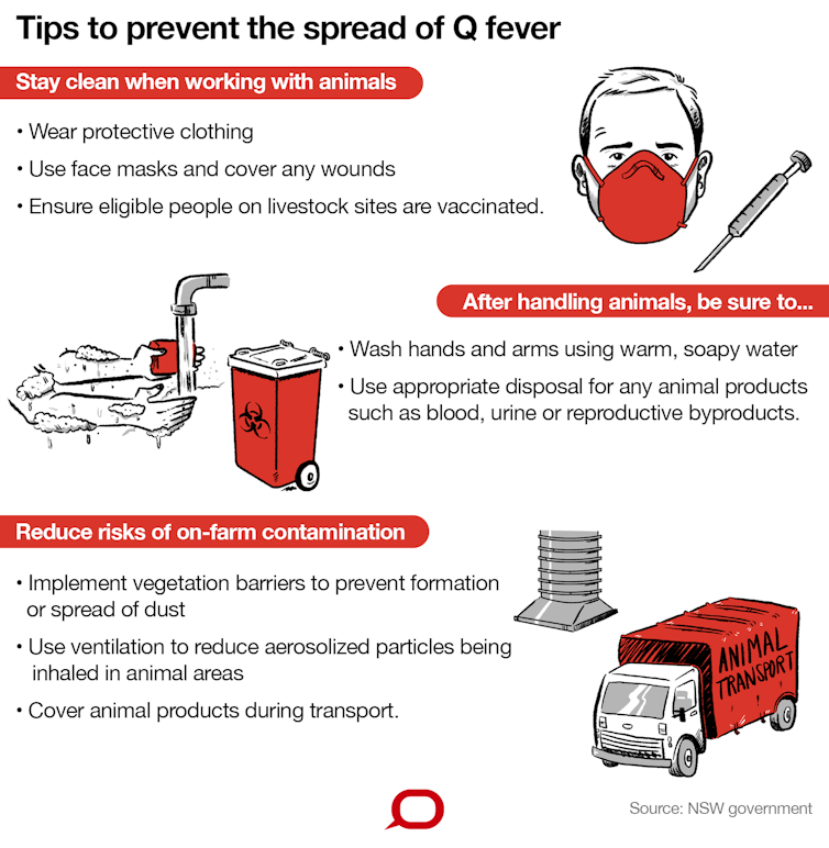
National guidelines for managing Q fever primarily recommend vaccination. The Q-VAX® vaccine has been in use since 1989. It’s safe and has an estimated success rate of 83–100%. However, people who have already been exposed to the bacteria are discouraged from having the vaccination, as they can develop a hypersensitive reaction to the vaccine. People aged under 15 years are also advised against the vaccine. Because the vaccine cannot be administered to everyone, people can take other steps to reduce risk. NSW Health recommends a series of precautions.
Author provided/The Conversation, CC BY-ND

What else can be done?

Vaccination for people in high-risk industries is effective to prevent Q fever infection, but must be administered well before people are actually at risk. Pre-testing requires both a skin test and blood test to ensure people who have already been exposed to the bacteria are not given the vaccine. This process takes one to two weeks before the vaccine can be administered, and it takes a further two weeks after vaccination to develop protection. This delay, along with the cost of vaccination, is sometimes seen as a barrier to its widespread use. Awareness of the vaccine may also be an issue. A recent study of Australians in metropolitan and regional centres found only 40% of people in groups for whom vaccination is recommended knew about the vaccine, and only 10% were vaccinated. We also need to better understand how transmission occurs in people who do not work with livestock (“non-traditional” exposure pathways) if we want to reduce Q fever rates.The Conversation Nicholas J Clark, Postdoctoral Fellow in Disease Ecology, The University of Queensland; Charles Caraguel, Senior lecturer, School of Animal and Veterinary Science, University of Adelaide; Jane Heller, Associate Professor in Veterinary Epidemiology and Public Health, Charles Sturt University; Ricardo J. Soares Magalhaes, Senior Lecturer Population Health & Biosecurity, The University of Queensland, and Simon Firestone, Academic, Veterinary Biosciences, University of Melbourne This article is republished from The Conversation under a Creative Commons license. Read the original article.
Dr Linda Calabresi

A decent eating program can keep you out of hospital, according to US research into the value of providing ready-to-consume meals to a select adult population. The retrospective cohort study involved just over 1000 participants, average age 53, almost 500 of whom were allocated to receive 10 meals a week, tailored to a recipient’s specific medical needs, for a period of just over two years. This group was then compared to a control group who had been matched for age and area of residence etc. Overall the study found the medically tailored meal delivery program was associated with approximately half the number of inpatient admissions over the duration of the study. Similarly, receiving the set meals was associated with significantly fewer admissions to skilled nursing facilities and a substantial reduction in health care costs. A pretty impressive result, yes? But before we go demanding an MBS item number for Meals on Wheels, even the researchers themselves advise caution in interpreting these results. Firstly, the study was not randomised. People who were allocated to receive the meal delivery intervention were generally more ill than the control patients – they were significantly more likely to have HIV, cancer and diabetes. “It is unlikely that similar results would be seen were the intervention applied to a healthier population, as the risk of admission or high health care costs, even in the absence of intervention would be substantially lower,” the study authors said in The Journal of the American Medical Association (JAMA) Internal Medicine.  And we don’t know whether it was the actual healthy food that made the difference, or whether it was the fact that they were getting their food for free thereby enabling the recipients to have more money for other things such as medications (remembering we are talking about the US health system here). Nonetheless, the study raises some valuable points. It is well-recognised that ‘following an appropriate diet is a cornerstone of maintaining health and managing illness.’ But this is often difficult for patients with complex medical conditions, especially if they are socioeconomically disadvantaged. As an accompanying editorial points out, much of the more recent focus has been on diet-related diseases and the health and economic burden they increasingly represent. Diseases such as diabetes, cardiovascular disease and obesity-related cancers have claimed much of the spotlight. But nutrition as a solution, and how we can use specific nutritional interventions to effectively manage a patient’s health care has been less well defined. “One obstacle has been demonstrating the efficacy and cost implications of specific nutritional interventions,” the editorial authors said. This JAMA study does that. Specifically, the researchers have shown that the provision of free, medically tailored meals at home is associated with reduced health care use and net cost savings. More importantly for Australians, the study supports the incorporation of nutrition into health care to improve patient health outcomes and keep vulnerable patients out of tertiary care. “Given their potential for significant health benefits and cost-savings, [medically tailored meals] may represent the tip of the spear for a national evolution toward such food-is-medicine approaches,” the editorial concludes.

Reference

Berkowitz SA, Terranova J, Randall L, Cranston K, Waters DB, Hsu J. Association Between Receipt of a Medically Tailored Meal Program and Health Care Use. JAMA Intern Med. Published online April 22, 2019. doi:10.1001/jamainternmed.2019.0198 Mozaffarian D, Mande J, Micha R. Food Is Medicine—The Promise and Challenges of Integrating Food and Nutrition Into Health Care. JAMA Intern Med. Published online April 22, 2019. doi:10.1001/jamainternmed.2019.0184
Emmanuel Stamatakis

As little as 20 minutes of exercise a day can offset a sedentary lifestyle. And that exercise can include walking the dog. People who spend much of their day sitting may need to move around less than we thought to counteract their sedentary lifestyle, new research shows. Our research, published today in the Journal of the American College of Cardiology, found about 20-40 minutes of physical activity a day seems to eliminate most health risks associated with sitting. That’s substantially lower than the one hour a day a previous study has found. We spend almost all our waking day sitting, standing, or moving. The health impact of each one of these can be complex. For example, too much standing can lead to lower back problems and even a higher risk of heart disease. But sitting for too long and not moving enough can harm our health. Then there are people who sit for many hours and also get in reasonable amounts of physical activity. For example, someone who has an office job but walks to and from work for 20 minutes each way and runs two to three times a week easily meets the recommended level of physical activity. While we know moving is better than sitting, what is far less clear is how much of a good thing (moving) can offset the harms of a bad thing (sitting). That’s what we wanted to find out in our study of almost 150,000 Australian middle-aged and older adults. We followed people enrolled in the 45 and Up Study for nearly nine years. We looked at links between sitting and physical activity with deaths from any cause, and deaths from cardiovascular disease such as heart disease and stroke, over that time. We then estimated what level of moderate-to-vigorous physical activity might offset the health risks of sitting. This kind of activity is strenuous enough to get you at least slightly out of breath if sustained for a few minutes. It includes brisk walking, cycling, playing sports or running.

What we found

People who did no physical activity and sat for more than eight hours a day had more than twice (107%) the risk of dying from cardiovascular disease compared to people who did at least one hour of physical activity and sat less than four hours a day (the “optimal group”). But it wasn’t enough just to sit less. People who did less than 150 minutes of physical activity a week and sat less than four hours a day still had a 44-60% higher risk of dying from cardiovascular disease than the optimal group. We also calculated the effect of replacing one hour of sitting with standing, walking, and moderate and vigorous physical activity. Among people who sit a lot (more than six hours a day) replacing one hour of sitting with equal amounts of moderate physical activity like strenuous gardening and housework, but not standing, was associated with a 20% reduction in dying from cardiovascular disease. Replacing one hour of sitting with one hour of vigorous activity such as swimming, aerobics and tennis, the benefits were much greater, with a 64% reduction in the risk of dying from cardiovascular disease.

What does it all mean?

The great news for people who sit a lot, including sedentary office workers, is that the amount of physical activity needed to offset the health risks of sitting risks was substantially lower than the one hour a day a previous study found. Even around 20-40 minutes of physical activity a day - the equivalent of meeting the physical activity guidelines of 150 to 300 minutes a week – seemed to eliminate most risks associated with sitting. For people who sat a lot, replacing sitting with vigorous physical activity was better than replacing it with moderate activity; and replacing sitting with moderate activity or walking was better than replacing it with standing.

What’s the take-home message?

Our study supports the idea that sitting and exercise are two sides of the same health “coin”. In other words, enough physical activity can offset the health risks of sitting. Should we worry about sitting too much? Yes, because sitting takes up valuable time we could spend moving. So too much sitting is an important part of the physical inactivity problem. We also know only a minority of adults get enough physical activity to offset the risks of sitting. For those who sit a lot, finding ways to reduce sitting would be a good start but it is not enough. The most important lifestyle change would be to look for or create opportunities to include physical activity into our daily routine whenever possible.

How to widen our activity ‘menu’

Not everyone has a supportive environment and the capacity to create opportunities to be active. For example, lack of time and physical activity being low on people’s list of priorities are the main reasons why inactive adults don’t exercise. Also, many do not have the motivation to power through a strenuous workout when they are juggling many other life challenges. There are no known remedies to a lack of time or low motivation. So, perhaps we need to add new approaches, beyond exercising and playing sport for leisure, to the “menu” of physical activity options. Incidental physical activity like active transportation – think walking fast or cycling part or all of the way to work – or taking stairs are great ways to become or stay active without taking much extra time.The Conversation Emmanuel Stamatakis, Professor of Physical Activity, Lifestyle, and Population Health, University of Sydney; Joanne Gale, Research Fellow Biostatistician, University of Sydney, and Melody Ding, Senior Research Fellow of Public Health, University of Sydney This article is republished from The Conversation under a Creative Commons license. Read the original article.
Dr Linda Calabresi

Benzos increase the risk of having a miscarriage in early pregnancy, regardless of whether you’re taking a short-acting one for insomnia or a longer-acting one for anxiety, Canadian researchers say. According to their large case-controlled study involving almost 450,000 pregnancies, benzodiazepine exposure in early pregnancy was associated with an 85% higher risk of spontaneous abortion compared to pregnancies where that class of drugs were not taken. And this increased risk remained the same, after a whole range of possible confounders had been adjusted for, including maternal mood and anxiety disorders. But this isn’t the new bit. Previous research, including both a UK population-based study and an Israeli prospective study had confirmed the link between benzos and spontaneous abortion. In Australia, benzodiazepines have been given a Category C rating in terms of safety in pregnancy. (Drugs owing to their pharmacological effects have caused or may be suspected of causing harmful effects on the human fetus or neonate without causing malformations.) “Benzodiazepines cross the placental barrier and accumulate in the fetal circulation at levels that are one to three times higher than the maternal serum levels”, the researchers explained. What hasn’t been known, to date, is whether this is a class effect or are some benzos worse than others. Well – according to this study published in JAMA Psychiatry – ‘the risk was similar among pregnancies exposed to short-acting …and long-acting benzodiazepines during early pregnancy.’ So it didn’t matter if you were prescribed long-acting clonazepam or short-acting lorazepam (interestingly the two most frequently prescribed benzos), the risk was more or less the same. “All benzodiazepine agents were independently associated with an increased risk of [spontaneous abortion],” the study authors said. In addition the study found the risk increased as the daily dose of benzodiazepines increased, suggesting a dose-response effect. So basically the more doses of benzos a pregnant woman takes, either in terms of strength or duration, the greater the risk she will miscarry. Overall, the researchers concluded that pregnant women should avoid taking benzodiazepines, and if they have to take them only take the lowest dose possible for shortest duration possible. “Alternative nonpharmacologic treatments exist and are recommended, but if benzodiazepines are needed, they should be prescribed for short durations,” they concluded.  

Reference

Sheehy O, Zhao JP, Bérard A. Association Between Incident Exposure to Benzodiazepines in Early Pregnancy and Risk of Spontaneous Abortion. JAMA Psychiatry. 2019 May 15. DOI: 10.1001/jamapsychiatry.2019.0963 [Epub ahead of print]  
Dr Linda Calabresi

Increasingly, pregnant women are heeding the warnings about the dangers of pertussis and getting vaccinated but the same does not appear to be happening with influenza protection. According to an Australian retrospective analysis, pertussis vaccination of pregnant women in Victoria increased from 38% in 2015 to 82% two years later. However, when they looked at rates of influenza vaccination the prevalence fluctuated according to the season but even so, the overall rate was only 39%. Looking first at the factors that appeared to influence whether a woman got vaccinated at all, the researchers found women who were older, who were having their first child, attended antenatal care earlier in the pregnancy and who were receiving GP-led care were more likely to receive immunisation (thumbs up for the GPs). On the negative side, the likelihood of vaccination was significantly lower in women born overseas, those who smoked during pregnancy and among Aboriginal and Torres Strait Islander women. Overall it appeared the more contact a pregnant woman had with the health system, especially if that contact was with health professionals who are well-versed in all things immunisation, ie GPs, the more likely it was that vaccination would be offered, accepted and delivered. The variation in coverage rates across different hospital-led organisations reflects the fact that immunisation for flu and pertussis has not yet become part of standard, best practice guidelines for routine antenatal care. “Fewer than half the respondents indicated that vaccines were always or usually administered during routine antenatal care,” they wrote. Following on from these general observations, researchers tried to determine why it was that vaccination coverage for pertussis rose so dramatically between 2015 and 2017, and why coverage for influenza prevention didn’t. “This may reflect continued promotion by state and national bodies of the importance of maternal pertussis vaccination, and increased awareness among pregnant women of the seriousness of pertussis in infants,” they said. By contrast, the researchers suggest that influenza is often believed to pose a greater health risk to the mother as opposed to the infant, and this along with concerns about the safety of the flu vaccine itself may, at least in part, explain the poor uptake of this vaccine. To improve this situation and increase rates of protection for Australian pregnant women and their children, the study authors had a number of recommendations. Most importantly they suggest we need to build vaccination against pertussis and influenza into the standard of care for all antenatal practices – be they hospital based, midwife-led or part of the GP antenatal shared care program. Basically we need to bring vaccination up and centre into our consciousness, so women get offered the vaccine and then ensure our systems have the capacity to be able to provide this vaccination as the opportunity arises. “Maternal vaccination should be embedded in all antenatal care pathways, and systems should be improved to increase the uptake of vaccination by pregnant women,” they conclude. Other recommendations included highlighting the benefits of vaccination to those groups of women most at risk such as women who smoke and Aboriginal and Torres Strait Islander women. But key to all the recommendations is making vaccination just part of routine care. As an accompanying editorial points out, “Embedding vaccination into standard pregnancy care, whether delivered by GPs, midwives or obstetricians, normalises the process, improves access to vaccination and reduces the risk of missing opportunities for vaccination.”  

References:

Rowe SL, Perrett KP, Morey R, Stephens N, Cowie BC, Nolan TM, et al. Influenza and pertussis vaccination of women during pregnancy in Victoria, 2015-2017. Med J Aust 2019 Jun 3; 210(10): 454-62. DOI: 10.5694/mja2.50125 Marshall HS, Amirthalingam G. Protecting pregnant women and their newborn from life-threatening infections. Med J Aust 2019 Jun 3; 210(10): 445-6. DOI: 10.5694/mja2.50174
Dr Linda Calabresi

Female patients who present with vulval pain or itch have usually put up with symptoms for some time before coming to the doctor. When it comes to this area of the female anatomy there is still a lot of ignorance and embarrassment in the world outside our surgeries. And while it is true you can find the answer to almost any question on the net, for these particular problems there’s a lot of dodgy information out there. So here’s an information source you can trust and recommend. Care down there (www.caredownthere.com.au) is a consumer-directed website written by respected health professionals that provides accurate, up-to-date and practical information and advice about all things vulval. From herpes to vulval sclerosis, the site covers the broad range of conditions that can affect women as well as providing some fundamental education about how to distinguish between normal anatomy and physiology and something going awry. The site was founded by Dr Gayle Fischer and Dr Jennifer Bradford who are well-known, well-respected members of the Australasian College of Dermatologists and the Royal Australian and the New Zealand College of Obstetricians and Gynaecologists. The content has been developed by a group of Australian health professionals with an interest and experience in vulval problems including dermatologists, gynaecologists, a pain management specialist, a sexual health physician, a psychologist and a pelvic floor physiotherapist. It is sponsored by Epiderm but exists as an independent resource. If you’re wanting to direct your female patients to a resource that is both comprehensive and authoritative, this really does fit the bill.   >> Access the resource here

Prof Mariano Barbacid

Pancreatic ductal adenocarcinoma (PDAC), the most common form of pancreatic cancer, is the third most common cause of death from cancer in the United States and the fifth most common in the United Kingdom. Deaths from PDAC outnumber those from breast cancer despite the significant difference in incidence rates. Late diagnosis and ineffective treatments are the most important reasons for these bleak statistics. PDAC is an aggressive and difficult malignancy to treat. Until now, the only chance for cure is the complete surgical removing of the tumor. Unfortunately, because PDAC is usually asymptomatic, by the time it is diagnosed 80% to 90% of patients have disease that is surgically incurable. PDAC thus remains one of the main biomedical challenges today due to its low survival rate – just 5% of patients are still alive five years after diagnosis. However, in recent decades a number of studies have shed light on the molecular mechanisms responsible for the initiation and progression of PDAC. Our recent research has shown that progress toward a cure is possible.

Ineffective treatments

The molecular mechanisms responsible for pancreatic cancer are complex. This is why recent advances in personalized medicine and immunotherapy (which helps the immune system fight cancer) have failed to improve the treatment of pancreatic cancer. This is mainly due to two characteristics:
  • 95% of these tumors are caused by mutations in KRAS oncogenes. Oncogenes are genes that, once mutated, are capable of inducing the transformation of a normal cell into a cancerous cell. KRAS is a gene that acts as an on/off switch. Normally, KRAS controls cell proliferation. When it is mutated, however, the cells start to grow uncontrollably and proliferate – a hallmark of cancer cells. So far, KRAS oncogenes have not been able to be targeted by drugs.
  • PDACs are surrounded by abundant fibrous connective tissue that grows around some tumor types. In the case of PDAC, this tissue forms a barrier that prevents cells that recognize and attack tumor cells, called cytotoxic T lymphocytes, from reaching the inside of the tumor mass and killing its cells. This renders immunotherapy treatments useless.
For these reasons, PDAC continues to be treated with drugs that destroy cancerous cells but can also destroy healthy ones. Options include Demcitabine, approved in 1997, and Nab-paclitaxel, a new paclitaxel-based formulation. Even if such a treatment is an option, it typically only extends the patients’ lives a few weeks, a marginal improvement at best. In recent years, however, a number of studies have shed light regarding the molecular mechanisms responsible for the initiation and progression of PDAC. Today we know that most of these tumors are caused by mutations in the KRAS oncogene. They lead to benign alterations that cause additional mutations in a range of tumor-suppressor genes, which usually repair DNA mistakes, slow down cellular division or tell cells when to die. Mutated cells can grow out of control, and in this context progress to malignant PDAC. While this process is relatively well known, it has not had an immediate impact on the development of new and more effective treatments.

In search of new strategies

Multiple strategies are currently being studied in an attempt to inhibit the growth of these tumors by blocking the growth of either the tumor cells or their surrounding “shielding” connective tissue. In our laboratory, we focused on blocking the signaling pathways that mediate the oncogenic activity of the initiating KRAS oncogenes. A decade ago, our lab decided to use genetically engineered mouse-tumor models capable of reproducing the natural history of human PDAC. We did this in order to analyze the therapeutic potential of the main components of the KRAS signaling pathways. These studies have unveiled the reason why the drugs tested so far have intolerable toxic effects, with mice dying within several weeks: they target some proteins that are essential for the dynamic state of equilibrium that is the condition of optimal functioning of the cells. This is called normal homeostasis. These crucial proteins are mainly kinases, enzymes that are able to modify how other molecules function. They play a critical and complex role in regulating cellular signaling and orchestrate processes such as hormone response and cell division. These results might explain why the KRAS-signaling inhibitors tested so far have failed in clinical trials. On the other hand, the removal of other signaling kinases did not have toxic side effects, but also had no impact on tumor development. Of the more than 15 kinases involved in the transmission of signals from the KRAS oncogene, only three displayed significant therapeutic benefits without causing unacceptable side effects. These are: RAF1, the epidermal growth factor receptor (EGFR) and CDK4.

It works! (in mice)

In initial studies, we observed that the elimination (via genetic manipulation) of the expression of some of these three kinases prevented the onset of PDAC caused by the KRAS oncogene. However, its elimination in animals with advanced tumors had no significant therapeutic effects. These results caused us to question whether it would be possible to eliminate more than one kinase simultaneously without increasing the toxic effects. As described in our recent work published in the journal Cancer Cell, the elimination of RAF1 and EGFR expression induced the complete regression of advanced PDACs in 50% of the mice. We are currently studying whether we can increase this by also eliminating CDK4. The analysis of the pancreas of animals in which we were no longer able to observe tumors by imaging techniques revealed the complete absence of lesions in two of them. Two mice showed some abnormal ducts, probably residual scarring from the tumor. The others had tumor micro-masses of one-thousandth the size of the original tumor. The study of these revealed the presence of tumor cells, in which the expression of the two targets, EGFR and RAF1, had not been completely eliminated, a common technical problem in this type of study. It is significant that these results were observed not only in mice. The inhibition of the expression of these two proteins in cells derived from nine out of ten human PDACs were also capable of blocking their proliferation in vivo when transplanted into immunosuppressed mice as well as in vitro cultures.

What now?

While these results have only been observed in a subset of mice for now, their importance lies in the fact that it is the first time that it has been possible to completely eliminate advanced PDAC tumors by eliminating a pharmacologically directed target. These observations are clearly important for the development of treatments based on the inhibition of RAF1 and EGFR, but they only represent a first step on a long, hard road ahead. First, it is important to identify the differences between the PDACs that respond to the combined elimination of RAF1 and EGFR and those that are resistant. As described in our work, the analysis of these two tumor types revealed that they are not active in the same way – more than 2,000 genes are expressed differently. Identifying additional targets in resistant tumors that do not increase treatment toxicity is not going to be an easy task. To continue our tests with genetically engineered mice, the immediate but no less difficult task is the development of specific RAF1 inhibitors. Indeed, we only currently have potent drugs against the second target, EGFR. In principle, there are four possible approaches:
  • Generate selective inhibitors for its kinase activity.
  • Generate inhibitors for its binding to the KRAS oncogene.
  • Generate inhibitors for its interaction with effector targets that transmit oncogenic signaling mediated by RAF1.
  • Degrade the RAF1 protein with drugs.
Designing inhibitors of the RAF1 kinase activity would seem to be the most affordable option, given the experience of the pharmaceutical industry in designing this molecule type. The problem resides in the fact that there are two other kinases of the same family, ARAF and BRAF, whose catalytic centers (the “active core” of the enzymes) are nearly identical. RAF1 kinase inhibitors are also targeting these other kinases, which causes collateral damage. The ones tested to date have caused high toxicities and the clinical trials had to be stopped. Continuing to develop effective molecules that are capable of blocking RAF1 activity in patients with PDAC will not be easy. It will surely take more time than we hope, but at least a road map has already been outlined that shows us how to keep moving forward. Created in 2007 to help accelerate and share scientific knowledge on key societal issues, the AXA Research Fund has been supporting nearly 600 projects around the world conducted by researchers from 54 countries. To learn more about this AXA Research Fund project, please visit the dedicated page. This article was translated from the original Spanish by Sara Crespo, Calamo & Cran.The Conversation Mariano Barbacid, profesor e investigador AXA-CNIO de Oncología Molecular, Centro Nacional de Investigaciones Oncológicas CNIO This article is republished from The Conversation under a Creative Commons license. Read the original article.
Dr Linda Calabresi

This is one of those resources you will wish you would have discovered earlier. Maternity Matters (https://maternity-matters.com.au/) is a website developed and largely authored by well-known Brisbane GP Dr Wendy Burton. As the name suggests it is reservoir of information related to pregnancy and is aimed chiefly at the general public but also provides a healthy serving of support materials for Australian health professionals as well. Dr Burton who is Chair of the Antenatal/Postnatal Specific Interest Group for the Royal Australian College of General Practitioners has compiled articles, interviews and links to important guidelines and similar reference materials on this site, that cover everything from pre-conception checklists to the post-natal issues such as vaccinations and return to exercise. This website is strongly based on evidence but is also very practical, addressing many of the more controversial and topical issues currently confronting the pregnant woman such as tongue-tie in infants and children’s risk of allergy. All the information is current and very Australian, with contributions from some very well-respected experts. As Dr Burton points out there is a lot of information out there on pregnancy and infant health - some of it very good, but, let’s face it, there’s a lot of dodgy stuff too – leaving women often confused and unnecessarily anxious. Asking patients not to Google is unlikely to be all that effective, but if patients can be directed to an authoritative, comprehensive site such as Maternity Matters you can feel confident that the information they receive will be reasonable and largely in keeping with what you, the qualified GP, would have said – if you were available at 3am when the patient was awake and seeking answers. Access the resource here

Dr Linda Calabresi

These days the first question most GPs get asked, after confirming a wanted first pregnancy is what does the woman need to take or eat, and, importantly what should she avoid. It gets tricky doesn’t it? If you avoided  everything that is said to potentially cause harm (according to the world wide web and social media) the pregnant woman will run a serious risk of malnutrition!  Much of the fear stems from the risk of contracting listeria – that surreptitious bacteria that can cause – very occasionally, severe infection in affected adults – but more importantly for the pregnant women can cause miscarriage, premature birth or stillbirth. You need some authoritative, credible information sources to fall back on when giving these vulnerable patients advice. Enter the Food Standards Australia and New Zealand website. On their website they have this fantastic resource: an information page entitled Listeria and food. Quite simply, it’s a precis of what women should and should not eat to keep them safe from this infection. To be fair this resource doesn’t help women keep the problem in perspective, as it doesn’t mention how rarely this condition affects pregnant women. But it gives very definitive advice – eat this – don’t eat that. The reality is, this could save the GP at least 15 minutes discussion time, and provide a ready reckoner for the woman negotiating the local café menu or shopping centre food halls. There is no debate, the pregnant woman, especially the first-time pregnant woman represents a very vulnerable, information-hungry demographic. Keep this site bookmarked and you won’t be sorry.   >> Access the resource here

Dr Smathi Chong

Herpes simplex virus (HSV) 1 and 2 are closely related to each other and more distantly related to Varicella Zoster virus (VZV), which causes Varicella (chicken pox) and Herpes Zoster (shingles). Traditionally HSV1 causes most oral herpes and HSV2 causes most genital herpes. But this is no longer so and has changed, probably due to more frequent oral sex. Figures from Clinipath 2017:
HSV Swab Origin HSV1 HSV2 VZV
Oral sites 93% 2% 5%
Genital/perineal sites 45% 50% 5%
HSV1 is frequently acquired in childhood and 75% of Australian adults would have had HSV1 by early adulthood. This would have been from oral contact with close friends and relatives who were shedding the virus, often asymptomatically. The classic “cold sores” are a blistering painful rash around the mouth Like other viruses in the Herpes family, this ‘lifelong’ infection can lay dormant and reactivate. The risk of reactivation and severe reactivation is higher in immunosuppressed individuals but in most people there is no readily identifiable reason for their reactivation. Stress is often blamed. Less common infections include:
  • HSV encephalitis (HSV1 in adults) and HSV meningitis (usually HSV2 in adults)
  • Conjunctivitis/keratitis – usually HSV1 or VZV (shingles affecting trigeminal nerve)
  • Herpetic whitlow – painful vesicles affecting the finger or thumb caused by HSV1 or 2

Genital Herpes

This causes most angst in patients as there is a social stigma. Approximately one in 7 of the general Australian adult population is seropositive to HSV2 but most are asymptomatic or subclinical. HSV Serology has a more limited role. Many clinicians (and patients) expect Herpes serology to be able to do more than it can! Test results may not answer many clinical or patients’ questions. A positive serology simply indicates a patient has been infected with HSV at some time in the past. It is not able to time the initial infection unless seroconversion (HSV IgG changing from negative to positive) can be demonstrated. In Herpes reactivation, the IgG would already be positive. Serology does not indicate the site of infection (e.g. oral or genital) although a strong positive HSV2 serology in the setting of painful genital lesions is likely to indicate genital herpes. Serology does not confirm whether symptoms are due to herpes. A positive PCR on a genital lesion would be more helpful. Positive serology is not able to tell if the person is infectious at the time of the test. HSV Serology or PCR would NOT be able to determine whether a person’s partner has been unfaithful! False positive (perhaps up to 5%) and false negative serology results can occur. Serology is often negative in acute primary herpes infection as HSV IgG can take a few weeks to a few months to become positive. Serology positivity may also decline over time. HSV IgM is no longer performed in most labs as they often throw up more confusion due to the non-specific nature of the test.

HSV in Pregnancy

HSV can cause severe neonatal infections including meningo-encephalitis, disseminated disease and even death. The highest risk is in symptomatic primary herpes infection of the birth canal/genital track. Herpes simplex serology may be more useful in the setting of pregnancy in patients with genital lesions suggestive of herpes to help risk stratify whether the episode is likely to be primary HSV. The highest risk would be PCR proven active genital lesions and negative serology. Treatment including anti-viral therapy and consideration of caesarean section may be discussed with the obstetrician. Management of the neonate with high risk of HSV should be handled by a neonatologist or paediatrician.

Treatment

These viruses may be treated with aciclovir, valaciclovir or famciclovir.
General Practice Pathology is a new regular column each authored by an Australian expert pathologist on a topic of particular relevance and interest to practising GPs. The authors provide this editorial, free of charge as part of an educational initiative developed and coordinated by Sonic Pathology.
Dr Linda Calabresi

If you work in a truly general general practice you will no doubt be familiar with this scenario. The patient on a NOAC who is going for a dental procedure, cataract operation or arthroscopy. Do they need to stop their NOAC? And if so, when and for how long? Often there are more than a couple of opinions out there, with a lot of different considerations to take into account.

Dr Linda Calabresi

Physical activity not only helps prevent depression, but should be considered an effective therapeutic option for patients who already have the condition, researchers say. According to a review published recently in Current Sports Medicine Reports, major depressive disorder is an exceedingly common, disabling condition with prevalence estimates ranging from 6% to 18% across different countries worldwide.